Photograph
Tick Removal
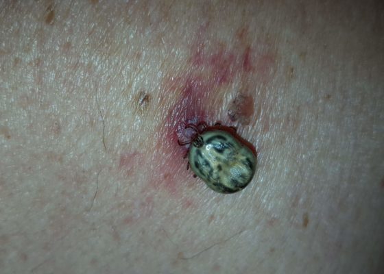
DOI: https://doi.org/10.21980/J8HK9HOn physical exam, an engorged tick was found attached to the patient’s left upper back. The underlying skin was nontender but mildly erythematous, without central clearing. The tick was gently removed with blunt angle forceps and sent for further analysis, which later revealed the specimen to be an American dog tick (Dermacentor variabilis).
Lightning Ground Current Injury: A Subtle Shocker
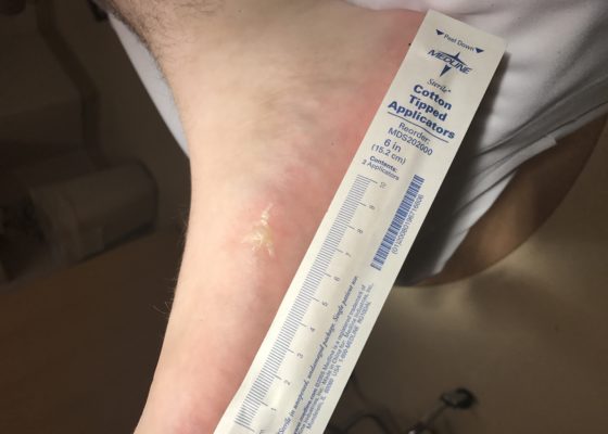
DOI: https://doi.org/10.21980/J8KD1CThe first photograph demonstrates a dendritic blister (Lichtenburg figure) on the medial aspect of his right foot where the ground current injury entered the patient's foot. Although no data exists regarding the sensitivity or specificity of Lichtenberg figures as skin findings, they are considered pathognomonic for lightning injuries and are not produced by alternating current or industrial electrical injuries. The second photograph demonstrates a 4 x 3 cm area of petechiae where the ground current injury exited the patient.
Facial Fracture Induced Periorbital Emphysema
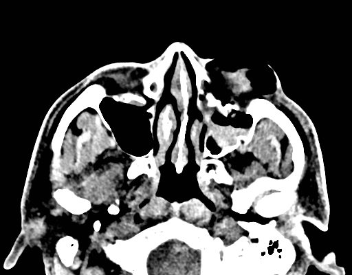
DOI: https://doi.org/10.21980/J8F05HPhysical exam showed marked left palpebral subcutaneous crepitus, as well as bulbar and palpebral conjunctival bulging. Visual acuity was normal with intact extraocular movements, and normal pupillary exam. Computed tomography (CT) imaging of the face was obtained and revealed multiple displaced fractures involving the left orbital floor and zygomatic arch associated with moderate periorbital and postseptal extraconal gas, resulting in orbital proptosis.
Corneal Rust Ring
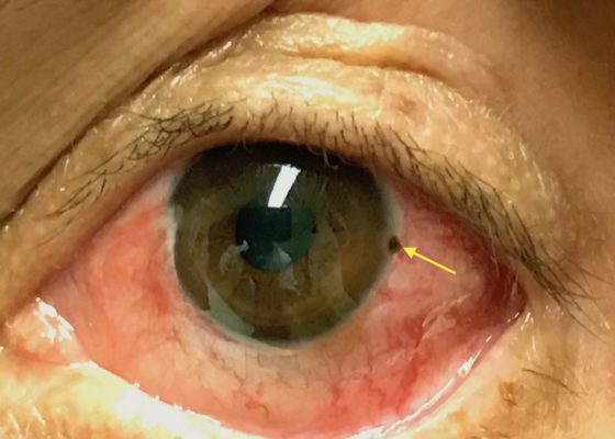
DOI: https://doi.org/10.21980/J8X067The photograph reveals a limbic metallic foreign body with a surrounding corneal rust ring (arrow) in the three o’clock position of the left cornea.
Cauliflower Ear Secondary to a Chronic Auricular Hematoma
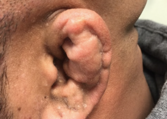
DOI: https://doi.org/10.21980/J8S63XOn exam, the patient has a gross deformity to the left pinna that was not painful to touch or fluctuant. Findings and history are consistent with cauliflower ear, secondary to a chronic auricular hematoma.
Various Degrees of Thermal Burns
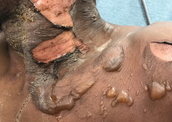
DOI: https://doi.org/10.21980/J8R91WOn exam,there is a large swath of skin with evidence of thermal injury involving the neck, shoulder, chest, and face, including damage to the ear, external nostril, and lips. Burns exhibit varying degrees of severity and total approximately 4.5% of the body surface area. Several areas are charred and insensate to pinprick. The left earlobe is partially burned off. Patient's airway is patent with no evidence of thermal injury or obstruction to the oropharynx or nasal vestibule.
Pemphigoid Gestationis
DOI: https://doi.org/10.21980/J8MG9DPhysical exam findings were significant for 1-3 cm diameter well-demarcated superficial ulcers on the patient’s abdomen and extremities, with mucosal sparing. Several small tense bullae were present on the bilateral inner thighs and numerous small reddish plaques were scattered over the patient’s back. Nikolsky’s sign was negative. No lymphadenopathy was noted.
Fight Bite with Tendon Laceration
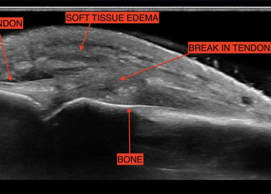
DOI: https://doi.org/10.21980/J8MP7QThe video shows a water bath ultrasound of the right 4th digit, demonstrating soft tissue swelling with a hypoechoic region along the tendon consistent with edema and tendon disruption (see video and annotated still image).

