Search By Topic
Found 26 Unique Results
Page 1 of 3
Page 1 of 3
Page 1 of 3
Retropharyngeal Abscess in an Adult Patient Presenting with Neck Fullness and Dysphagia: A Case Report
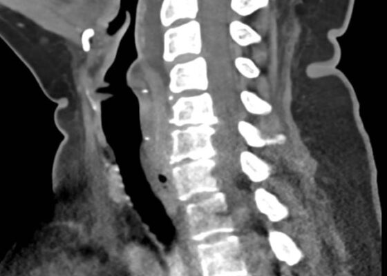
DOI: https://doi.org/10.21980/J8M36GContrast-enhanced CT soft tissue of the neck showed evidence of a prevertebral/retropharyngeal fluid collection, extending from the odontoid tip to the inferior C4 vertebral body margin, measuring 5.4 x 1.0 x 3.3 centimeters (cm) in size (yellow lines) without gross airway narrowing.
The Advantage of Using Video Laryngoscope in Puncture and Incisional Drainage of Peritonsillar Abscess: A Case Report
DOI: https://doi.org/10.21980/J8G935Incision of the peritonsillar abscess was performed with the assistance of the C-MAC video laryngoscope which provided a clear, illuminated, and unobstructed view of the incision site. Local anesthesia with 1% xylocaine was administered, and the abscess was incised with a scalpel and drained with a forceps.
A Man with Sore Throat—A Case Report
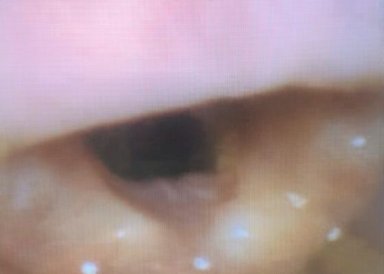
DOI: https://doi.org/10.21980/J8MH0BVideo laryngoscopy of the upper airway was performed two days after initial burn injury. The images obtained demonstrated laryngeal edema and inflammation near the epiglottis. The dot identifies the epiglottis and the asterix identifies the area of moderate thermal burns. Imaging also demonstrated adequate patency of airway and ruled out the need for intubation at that time.
Evaluation of ACE-inhibitor Induced Laryngeal Edema Using Fiberoptic Scope: A Case Report
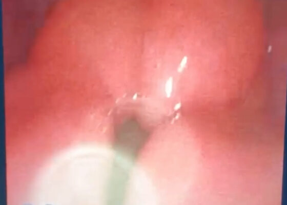
DOI: https://doi.org/10.21980/J83P9TPhysical exam was initially significant for swelling isolated to the right sided cheek and upper lip. There was no edema to lower lip, uvular swelling, or swelling to the submandibular space. She was speaking full sentences and did not endorse any voice changes. Initial vital signs were as follows: BP 125/77, HR 74, RR 16, and oxygen saturation of 100% on room air. Approximately 40 minutes later, after 125 mg solumedrol intravenous (IV) and 50mg diphenhydramine by mouth, swelling had spread to the entire upper lip and the patient reported spreading to her jaw (Photo 1). Although no jaw or submandibular edema was appreciated on physical exam, a flexible fiberoptic laryngoscope was used to evaluate the patient’s airways given worsening symptoms. Viscous lidocaine was applied intranasally five minutes prior to the procedure. The patient was positioned in a seated position on the stretcher. A flexible fiberoptic laryngoscope was then inserted through the nares and advanced slowly. Laryngoscopy showed diffuse edema of the epiglottis, arytenoids, and ventricular folds (see photos 2-4). Vital signs and respiratory status remained stable both during and after the procedure.
Peritonsillar Abscess Simulator: A Low-Cost, High-Fidelity Trainer
DOI: https://doi.org/10.21980/J85M0BBy the end of this training session, learners will be able to: 1) locate the abscess, 2) perform needle aspiration, and 3) develop dexterity in maneuvering instruments in the small three-dimensional confines of the oral cavity without causing injury to local structures.
A Case Report of Epiglottitis in an Adult Patient
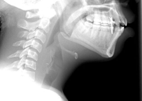
DOI: https://doi.org/10.21980/J8QM09At the time of presentation to the ED, laboratory results were significant for leukocytosis to 11.8 x 10⁹ white blood cells/L and a partial pressure of carbon dioxide of 52 mmHg on venous blood gas. Computed tomography (CT) of the soft tissue of the neck with contrast showed edematous swelling of the epiglottis and aryepiglottic fold with internal foci of gas (blue arrow) and partial effacement of the laryngopharyngeal airway and scattered cervical lymph nodes bilaterally (Figure 1). Findings were consistent with epiglottitis containing nonspecific air. Additionally, the pathognomonic “thumbprint sign” (yellow arrow) was found on lateral x-ray of the neck (Figure 2). The CT findings as shown in figure 3 illustrate lateral view of the swelling of the epiglottis, gas, and blockage of the airway.
A Low-Cost Facial and Dental Nerve Regional Anesthesia Task Trainer
DOI: https://doi.org/10.21980/J8RP9QBy the end of this educational session, learners should be able to: 1) describe and identify relevant anatomy for supra-orbital, infra-orbital, mental, and inferior alveolar nerves and 2) successfully demonstrate supra-orbital, infra-orbital, mental, and inferior alveolar nerve blocks using a partial task trainer.
Auricular Perichondritis after a “High Ear Piercing:” A Case Report
DOI: https://doi.org/10.21980/J8WH16On physical examination, there was erythema, swelling, warmth, and general exquisite tenderness of the superior aspect of the left pinna (the outer ear) but excluding the ear canal, lobe, tragus, and crus. There was no facial involvement. There was no fluctuance about the ear and no drainage of fluid. The preauricular lymph nodes were enlarged and tender, but the anterior cervical lymph nodes were not tender. There was no mastoid tenderness, protrusion of the ear, or interruption of the postauricular crease.
Peritonsillar Abscess Model for Ultrasound Diagnosis Using Inexpensive Materials
DOI: https://doi.org/10.21980/J86G9PBy the end of this instructional session learners should be able to: 1) identify and discuss the indications, contraindications, and complications associated with peritonsillar abscesses, 2) properly identify and measure a PTA through ultrasound, and 3) competently perform ultrasound-guided peritonsillar abscess drainage on a simulator and remove fluid.
Case Report: Acute Supraglottitis
DOI: https://doi.org/10.21980/J8006VOn arrival, radiographs of the neck soft tissues were obtained, which showed a markedly enlarged epiglottic shadow (red arrow) concerning for epiglottitis. A computed tomography scan of the neck soft tissues with contrast was then obtained which revealed edematous mucosal thickening of the oropharynx (blue arrow) and supraglottic larynx (green arrow) including the epiglottis (purple arrow) concerning for acute infectious pharyngitis and supraglottic laryngitis with severe narrowing of the supraglottic laryngeal lumen, as well as associated extensive inflammation and edema of the superficial and deep left neck spaces. The patient’s white blood cell count was elevated to 25.7x10⁹/L with 87% neutrophils. Her rapid strep test was positive. Otolaryngology was consulted and performed a bedside flexible laryngoscopy which showed significant edema of the epiglottis (orange arrow), vocal cords (white arrow), and arytenoids (black arrow), left greater than right. Based on the findings and concern for impending respiratory failure, the patient received an awake fiberoptic intubation by anesthesia at the bedside.


