Visual EM
Ovarian Teratoma
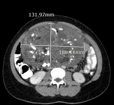
DOI: https://doi.org/10.21980/J8934XThe CT scan with oral contrast in the emergency department revealed a large heterogeneous abdominopelvic mass measuring 13.2 x 18.8 x 23.1 cm (see white lines), suggestive of an ovarian teratoma from the right ovary. This mass included fat, fluid, calcifications (see yellow arrows), and enhancing soft tissue components. The teratoma resulted in mass effect upon large and small bowel loops (see blue highlighted areas), inferior vena cava (IVC), distal aorta (see red highlighted area) and right common iliac artery. A small volume of ascites was also observed. There was no evidence of bowel obstruction, vascular occlusion or other significant emergent finding. Additionally, transabdominal and transvaginal ultrasound images were obtained. The transabdominal image visualized the abdominopelvic mass (see four yellow stars). The transvaginal image visualized a cross section of the teratoma (see four red stars) in relation to the bladder (see four blue stars).
Warm & Blue: A Case of Methemoglobinemia
DOI: https://doi.org/10.21980/J8591MThe patient hadperioral cyanosis, blue coloration around her mouth, but the rest of the skin on her face appeared normal. She also had acrocyanosis to bilateral hands that can be seen in the image. The patient has a tan complexion up to the level of her wrists, but the palms of her hands are pale and cyanotic.
Intramural Hematoma with Type B Aortic Dissection
DOI: https://doi.org/10.21980/J81M03Computed tomography angiography of the chest and abdomen revealed a 9.5 cm thoracoabdominal aneurysm (red outline) with intramural hematoma (yellow shading) and large left pleural effusion versus hemothorax with old blood (blue shading).
Radiolucent Foreign Body Seen on Point-of-Care Ultrasound but not on X-ray
DOI: https://doi.org/10.21980/J8WS77X-rays of the foot were obtained and no radiopaque foreign body was visualized. Due to high clinical suspicion for retained foreign body, a point-of-care ultrasound was performed by applying a high-frequency linear probe at the area of discomfort. In the long axis an ovoid focus of hypoechogenicity (orange outline) is visualized. Within this finding there is a linear focus (yellow line) of increased echogenicity measuring 1 mm in diameter and 1 cm in length. On short axis view, a rectangle focus (green dot) demonstrating shadowing (blue highlight) is seen.
Open Globe with Intraocular Foreign Body
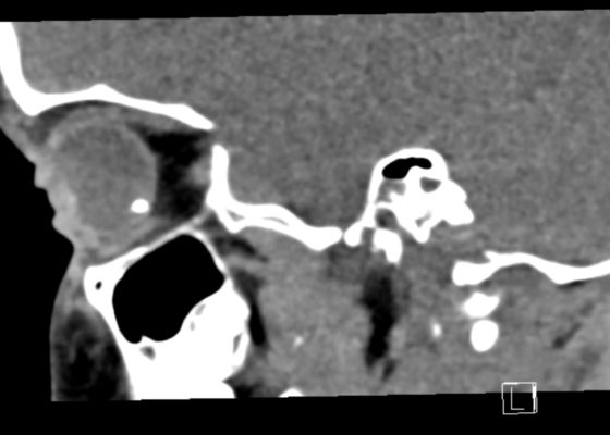
DOI: https://doi.org/10.21980/J8S348On physical exam, his extraocular movements were intact. The right anterior chamber appeared cloudy, particularly nasal to the pupil. The conjunctiva of the right eye was injected. The right pupil was 3 mm and sluggishly reactive and appeared slightly irregular (see yellow arrow). Of note, the right eye also had a 1 mm hypopyon, indicating inflammation of the anterior chamber, which was visible on slit lamp examination (not pictured). There was no fluorescein uptake or Seidel sign. His visual acuity was 20/60 OD (right eye) and 20/20 OS (left eye).
Hypopyon
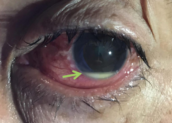
DOI: https://doi.org/10.21980/J8N92BPhysical examination of the left eye revealed a hypopyon (green arrow) – which is a layered white to yellow sediment in front of the inferior aspect of the iris associated with scleral injection and chemosis. Extraocular movements were intact bilaterally and pain did not worsen with extraocular movement. The pupil was poorly reactive to direct light and only hand movement could be perceived. The intraocular pressure was 14 mmHg. Slit lamp exam demonstrated a dense cataract. Bedside ocular ultrasound demonstrated vitreous opacities concerning for possible intraocular foreign bodies.
Tick Removal
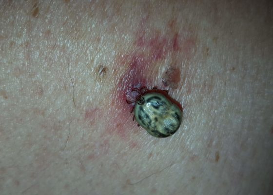
DOI: https://doi.org/10.21980/J8HK9HOn physical exam, an engorged tick was found attached to the patient’s left upper back. The underlying skin was nontender but mildly erythematous, without central clearing. The tick was gently removed with blunt angle forceps and sent for further analysis, which later revealed the specimen to be an American dog tick (Dermacentor variabilis).
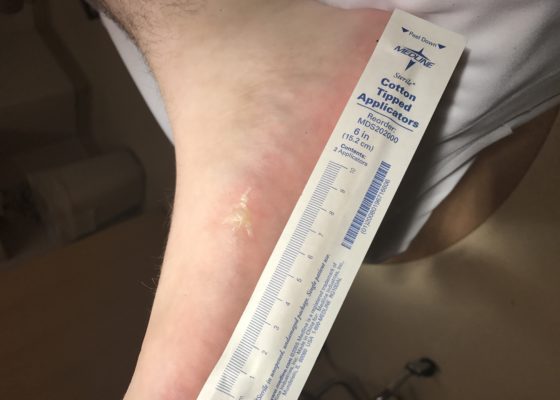
Lightning Ground Current Injury: A Subtle Shocker
DOI: https://doi.org/10.21980/J8KD1CThe first photograph demonstrates a dendritic blister (Lichtenburg figure) on the medial aspect of his right foot where the ground current injury entered the patient's foot. Although no data exists regarding the sensitivity or specificity of Lichtenberg figures as skin findings, they are considered pathognomonic for lightning injuries and are not produced by alternating current or industrial electrical injuries. The second photograph demonstrates a 4 x 3 cm area of petechiae where the ground current injury exited the patient.

