Acromioclavicular joint separation
History of present illness:
A 30-year-old male was brought in by ambulance to the emergency department as a trauma activation after a motorcycle accident. The patient was the helmeted rider of a motorcycle traveling at an unknown speed when he lost control and was thrown off his vehicle. He denied loss of consciousness, nausea, or vomiting. The patient’s vital signs were stable and his only complaint was pain around his left shoulder. On exam, the patient had a prominent left clavicle without skin compromise. He had adequate range of motion in the left shoulder with moderate pain, and his left upper extremity was neurovascularly intact.
Significant findings:
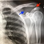
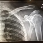
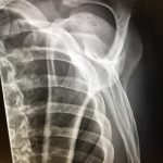
Plain films of the left shoulder showed elevation of the left clavicle above the acromion. There was an increase in the acromioclavicular (AC) and coracoclavicular (CC) distances (increased joint distances marked with red and blue arrows, respectively). A normal AC joint measures 1-3 mm whereas a normal CC distance measures 11-13 mm.1 The injury was classified as a Rockwood type III AC joint separation.
Discussion:
The AC joint is a synovial joint between an oval facet on the acromion and a similar facet on the distal end of the clavicle. Horizontal stability is provided by the AC joint while axial stability is provided by the CC joint.2,3 AC joint injuries account for about 9%-12% of shoulder girdle injuries, and the most common mechanism is direct trauma.4,5
Initial evaluation with imaging includes plain films with three views: the anterior-posterior (AP) view with the shoulder in internal and external rotation as well as an axillary, or scapula-Y view (sensitivity 40%, specificity 90% for all films).6,7 AC joint injuries are classified by the Rockwood system.8 Type I involves a sprain or incomplete tear of the AC ligaments with an intact CC ligament. The AC joint appears normal on X-ray, but can become widened with stress, achieved by having the patient hold a 10-15 pound weight from each forearm.1,9 Type II injuries involve a torn AC ligament, disrupting the AC joint. The AC joint appears widened on radiographs.1,9 The AC and CC ligaments are disrupted in type III injuries with an increased CC distance of 25%-100% on plain films.1,10 In addition to torn AC and CC ligaments, the clavicle is posteriorly displaced in a type IV injury. Because the AP film may not reveal the posterior displacement of the clavicle, the axillary view is vital for correct classification of type IV injuries.1 A type V injury involves disruption of the AC and CC ligaments as well as torn muscle attachments of the trapezius and deltoid on the clavicle and scapula, leading to greater AC joint displacement on radiographs. The CC distance appears 100%-300% greater than normal.1,10 Type VI injuries are caused by a direct blow to the superior surface of the clavicle resulting in inferior displacement. On X-ray, the lateral end of the clavicle is inferior to the acromion and coracoid processes in Type VI injuries.1
Treatment for types I, II, and III is conservative, which consist of analgesics, non-steroidal anti-inflammatory drugs (NSAIDs), and progressive range of motion exercises.11 Patients with type III injuries who fail conservative management are referred for operative repair.11,12 Type IV, V, and VI injuries require orthopedic consult and surgical management. Type IV injuries are treated with closed reduction to type III, followed by the treatment regimen for type III injuries.11 Like type III injuries, if conservative management of type IV injuries is unsuccessful, surgical management should follow.11
Topics:
Acromioclavicular joint, Rockwood classification, shoulder injury.
References:
- Ha AS, Petscavage-Thomas JM, Tagoylo GH. Acromioclavicular joint: the other joint in the shoulder. Am J Roentgenol. 2014;202(2):375-85. doi: 10.2214/AJR.13.11460
- Drake RL, Vogl W, Mitchell AW. Gray’s Anatomy for Students. Philadelphia PA: Elsevier; 2014;7:705-710.
- Saccomanno MF, Deieso C, Milano G. Acromioclavicular joint instability: anatomy, biomechanics and evaluation. 2014;2(2):87-92.
- Warth RJ, Martetschläger F, Gaskill TR, Millett PJ. Acromioclavicular joint separations. Curr Rev Musculoskelet Med. 2013;6(1):71-8. doi: 10.1007/s12178-012-9144-9
- Chillemi C, Franceschini V, Dei giudici L, et al. Epidemiology of isolated acromioclavicular joint dislocation. Emerg Med Int. 2013;2013:171609. doi: 10.1155/2013/171609
- Pogorzelski J, Beitzel K, Ranuccio F, et al. The acutely injured acromioclavicular joint – which imaging modalities should be used for accurate diagnosis? A systematic review. BMC Musculoskelet Disord. 2017;18(1):515. doi: 10.1186/s12891-017-1864-y
- Walton J, Mahajan S, Paxinos A, et al. Diagnostic values of tests for acromioclavicular joint pain. J Bone Joint Surg Am. 2004;86-A(4):807-12.
- Gastaud O, Raynier JL, Duparc F, et al. Reliability of radiographic measurements for acromioclavicular joint separations. Orthop Traumatol Surg Res. 2015;101(8 Suppl): S291-5. doi: 10.1016/j.otsr.2015.09.010
- Antonio GE, Cho JH, Chung CB, Trudell DJ, Resnick D. Pictorial essay. MR imaging appearance and classification of acromioclavicular joint injury. Am J Roentgenol. 2003;180(4):1103-10. doi: 10.1148/rg.282075714
- Alyas F, Curtis M, Speed C, Saifuddin A, Connell D. MR imaging appearances of acromioclavicular joint dislocation. Radiographics. 2008;28(2):463-79. doi: 10.1148/rg.282075714
- Montellese P, Dancy T. The acromioclavicular joint. Prim Care. 2004;31(4):857-66. doi: 10.1016/j.pop.2004.07.011
- Spencer EE. Treatment of grade III acromioclavicular joint injuries: a systematic review. Clin Orthop Relat Res. 2007;455:38-44. doi: 10.1097/BLO.0b013e318030df83


