Large Ventral Hernia
History of present illness:
A 46-year-old female presented to the emergency department (ED) with diffuse abdominal pain and three days of poor oral intake associated with non-bilious, non-bloody vomiting. Initial vital signs consisted of a mild resting tachycardia of 111/minute with a temperature of 38.0°C. On examination, the patient had a large pannus extending to the knees, which contained a hernia. She was tender in this region on examination. Laboratory values included normal serum chemistries and mild leukocytosis of 12.2. The patient reports that her abdomen had been enlarging over the previous 8 years but had not been painful until 3 days prior to presentation. The patient had no associated fever, chills, diarrhea, constipation, chest pain or shortness of breath.
Significant findings:
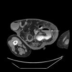
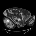
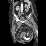
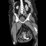
Computed tomography (CT) scan with intravenous (IV) contrast of the abdomen and pelvis demonstrated a large pannus containing a ventral hernia with abdominal contents extending below the knees (white circle), elongation of mesenteric vessels to accommodate abdominal contents outside of the abdomen (white arrow) and air fluid levels (white arrow) indicating a small bowel obstruction.
Discussion:
Hernias are a common chief complaint seen in the emergency department. The estimated lifetime risk of a spontaneous abdominal hernia is 5%.1 The most common type of hernia is inguinal while the next most common type of hernia is femoral, which are more common in women.1 Ventral hernias can be epigastric, incisional, or primary abdominal. An asymptomatic, reducible hernia can be followed up as outpatient with a general surgeon for elective repair.2 Hernias become problematic when they are either incarcerated or strangulated. A hernia is incarcerated when the hernia is irreducible and strangulated when its blood supply is compromised. A complicated hernia, especially strangulated, can have a mortality of greater than 50%.1
It is key to perform a thorough history and physical exam and to obtain appropriate imaging. Contrasted CT can best define hernias with a sensitivity and specificity of 83% and 67%-83%, respectively.2 One can also use point-of-care ultrasound to appreciate if there is an associated small bowel obstruction via the to-and-fro sign of peristalsis going both directions.2,3 The sensitivity of ultrasound for detection of a hernia is 92.7% with a specificity of 81.5%. If a hernia is incarcerated or strangulated, emergent surgical consultation is warranted for operation and reduction to prevent necrosis of the bowel wall and subsequent rupture.4
In this case, general surgery was consulted, and the patient was admitted to their service. After admission, she developed hypotension, fever, and metabolic acidosis. A repeat CT scan revealed thickened small bowel loops with extraluminal air, concerning for perforation. The patient was taken to the operating room emergently for exploratory laparotomy, where she was found to have murky peritoneal fluid and necrotic bowel with extensive adhesions. She subsequently required multiple operations for continued hemodynamic instability, intraabdominal necrosis and washout. The patient received a split thickness skin graft for abdominal closure and continues to receive feeds via Dobhoff tube secondary to amount of bowel resected.
Topics:
Hernia, small bowel obstruction.
References:
- Murphy KP, Owen OJ, Maher MM. Adult abdominal hernias. AJM Am J Roentgenol. 2014;202(6):W506-W511 doi: 10.2214/AJR.13.12071
- van den Berg JC, de Valois JC, Go PM, Rosenbusch G. Detection of groin hernia with physical examination, ultrasound, and MRI compared with laparoscopic findings. Invest Radiol. 1999;34(17) 739-743.
- Emby DJ, Georges A. CT Technique for suspected anterior abdominal wall hernia. AJM Am J Roentgenol.2003;181(2):431-433. doi: 10.2214/ajr.181.2.1810431
- Liang MK, Holihan JL, Itani K, et al. Ventral hernia management: expert consensus guided by systematic review. Ann Surg. 2017;265(1):80–89. doi: 10.1097/SLA.0000000000001701
- Brooks DC, Rosen M, Chen W. Overview of abdominal wall hernias in adults. Post TW, ed. UpToDate. Waltham, MA: UpToDate Inc. uptodate.com/contents/overview-of-abdominal-wall-hernias-in-adults. Published January 21, 2017. Accessed April, 4 2018.

