• Registration
  • Login
JETem
  • Home
  • About
    • Aim and Scope
    • Our Team
    • Editorial Board
    • FAQ
  • Issues
    • Current Issue
    • Ahead of Print
    • Past Issues
  • Visual EM
    • Latest Visual EM
    • Search Visual EM
    • Thumbnail Library
  • For Authors
    • Instructions for Authors
    • Submit to JETem
    • Photo Consent
    • Policies
      • Peer Review Policy
      • Copyright Policy
      • Editorial Policy, Ethics and Responsibilities
      • Conflicts of Interest & Informed Consent
      • Open Access Policy
  • For Reviewers
    • Instructions for JETem Reviewers
    • Interested in Being a JETem Reviewer?
  • Topic
    • Abdominal / Gastroenterology
    • Administration
    • Board Review
    • Cardiology / Vascular
    • Clinical Informatics, Telehealth and Technology
    • Dermatology
    • EMS
    • Endocrine
    • ENT
    • Faculty Development
    • Genitourinary
    • Geriatrics
    • Hematology / Oncology
    • Infectious Disease
    • Miscellaneous
    • Neurology
    • Ob / Gyn
    • Ophthalmology
    • Orthopedics
    • Pediatrics
    • Pharmacology
    • Procedures
    • Psychiatry
    • Renal / Electrolytes
    • Respiratory
    • Social Determinants of Health
    • Toxicology
    • Trauma
    • Ultrasound
    • Urology
    • Wellness
    • Wilderness
  • Modality
    • Curricula
    • Innovations
    • Lectures
    • Oral Boards
      • Structured Interview
      • Communication Case
    • Podcasts
    • Simulation
    • Small Group Learning
    • Team Based Learning
    • Visual EM
  • Contact Us

Pediatric Pulmonary Abscess

Kyle Barbour* and Jonathan Peña, MD*

*University of California, Irvine, Department of Emergency Medicine, Orange, CA

Correspondence should be addressed to Jonathan Peña, MD at jdpena@uci.edu 

DOI: https://doi.org/10.21980/J83S6QIssue 3:2
RespiratoryInfectious DiseaseVisual EM
No ratings yet.
Creative Commons images

History of present illness:

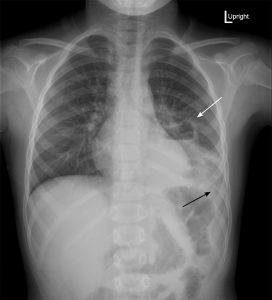
A 6-year-old previously healthy male presented to the emergency department with three days of left upper quadrant abdominal pain. Family endorsed one week of fevers, cough productive of yellow sputum, and non-bilious, non-bloody emesis. He denied shortness of breath and chest pain. On exam, the patient was febrile with otherwise normal vital signs. He had diffuse tenderness to his abdomen but clear lungs. Laboratory studies revealed leukocytosis to 25,000/mm3 with a left shift.

Significant findings:

Upright posterior-anterior plain chest films show a left lower lobe consolidation with an air-fluid level and a single septation consistent with a pulmonary abscess (white arrows). A small left pleural effusion was also present, seen as blunting of the left costophrenic angle and obscuration of the left hemidiaphragm (black arrows).

Discussion:

Pediatric pulmonary abscesses are rare, most commonly caused by aspiration, and the majority consequently arise in dependent portions of the lung.1 The most common pathogens in children are Streptococcus pneumoniaeand Staphylococcus aureus.1 Immunocompromised patients and those with existing pulmonary disease more commonly contract Pseudomonas aeruginosaor Bacteroides, and fungal pathogens are possible.1 Common symptoms include tachypnea, fever, and cough. Imaging is necessary to distinguish pulmonary abscesses from pneumonia, empyema, pneumatocele, and other etiologies.

Plain film radiography may miss up to 18% of pulmonary abscesses yet is often the first modality to visualize an intrathoracic abnormality.2 If seen, pulmonary abscesses most often appear as consolidations with air-fluid levels. Generally, pulmonary abscesses are round with irregular, thick walls, whereas empyemas are elliptical with smooth, thin walls.3 However, these characteristics cannot definitively distinguish these processes.2 Advantages of plain films include being low cost and easily obtained. Computed tomography (CT) of the chest with contrast has 100% sensitivity for pulmonary abscess and empyema but is more expensive and less frequently available.

Most patients require admission and parenteral antibiotics covering Staphylococcus, Streptococcus, anaerobes, gram-negative bacilli, and in selected cases, Pseudomonas. Combined ceftriaxone and clindamycin is generally effective.1,5,6 With treatment, mortality is less than 5%, significantly better than for adults.1

Topics:

Pulmonary abscess, pediatrics, radiograph, air-fluid level, infection, empyema, lung.

References:

  1. Patradoon-Ho P, Fitzgerald DA. Lung abscess in children. Paediatr Respir Rev. 2007;8(1):77-84. doi: 10.1016/j.prrv.2006.10.002
  2. Stark DD, Federle MP, Goodman PC, Podrasky AE, Webb WR. Differentiating lung abscess and empyema: radiography and computed tomography. AJR Am J Roentgenol.1983;141(1):163-167. doi: 10.2214/ajr.141.1.163.
  3. Walker CM, Abbott GF, Greene RE, Shepard JO, Vummidi D, Digumarthy SR. Imaging pulmonary infection: classic signs and patterns. AJR Am J Roentgenol. 2014;202(3):479-492. doi: 10.2214/AJR.13.11463
  4. Groskin SA, Panicek DM, Ewing DK, Rivera F, Math K, Teixeira J, et al. Bacterial lung abscess: a review of the radiographic and clinical features of 50 cases. J Thorac Imaging. 1991;6(3):62-67. doi: 10.1097/00005382-199107000-00008
  5. Puligandla PS, Laberge JM. Respiratory infections: pneumonia, lung abscess, and empyema. Sem Pediatr Surg. 2008;17(1):42-52. doi: 10.1053/j.sempedsurg.2007.10.007
  6. Tan TQ, Seilheimer DK, Kaplan SL. Pediatric lung abscess: clinical management and outcome. Pediat Infect Dis J. 1995;14(1):51-55. doi: 10.1097/00006454-199501000-00011
Icon

Pediatric Pulmonary Abscess - Case Report

1 file(s) 443 KB
Download
Icon

Pediatric Pulmonary Abscess - Images

1 file(s) 974.42 KB
Download
Issue 3:2X-Ray

Reviews:

No ratings yet.

Please rate this


Creative Commons images

Scaphoid Fracture

13 Apr, 18
Creative Commons images

Torsades de Pointes

13 Apr, 18

JETem is an online, open access, peer-reviewed, journal-repository for EM educators. We are PMC Indexed.

Most Viewed

  • The Silent Saboteur: Teaching the Clinical Implications of Occult Hypoxemia & Social Determinants of Health via a Pulmonary Embolism Case
  • Diabetic Ketoacidosis and Necrotizing Soft Tissue Infection
  • My Broken Heart
  • Stabilization of Cardiogenic Shock for Critical Care Transport, a Simulation
  • Innovative Ultrasound-Guided Erector Spinae Plane Nerve Block Model for Training Emergency Medicine Physicians

Visit Our Collaborators

Creative Commons Licence
This work is licensed under a Creative Commons Attribution 4.0 International License.

About

Education

Learners should benefit from active learning. JETem accepts submissions of team-based learning, small group learning, simulation, podcasts, lectures, innovations, curricula, question sets, and visualEM.

Scholarship

We believe educators should advance through the scholarship of their educational work. JETem gives educators the opportunity to publish scholarly academic work so that it may be widely distributed, thereby increasing the significance of their results.

Links

  • Home
  • Aim and Scope
  • Current Issue
  • For Reviewers
  • Instructions for Authors
  • Contact Us

Newsletter

Sign up to receive updates from JETem regarding newly published issues and findings.

Copyright Creative Commons Attribution 4.0 International