Visual EM
Case Report of Spontaneous Thyroid Hemorrhage Following LMA Insertion
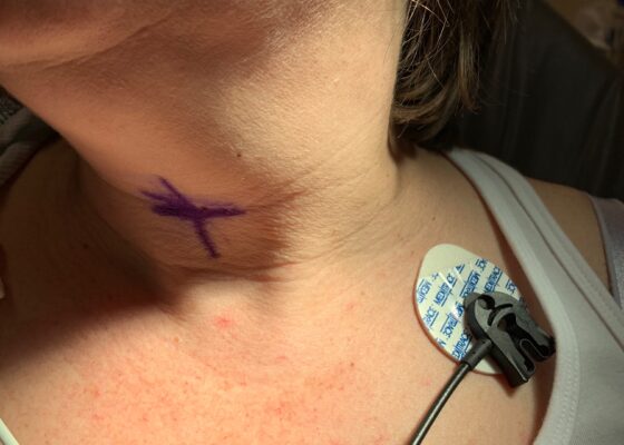
DOI: https://doi.org/10.21980/J8XP8WTwo photographs of patients neck, both showcasing no obvious erythema, bruising, or swelling which is noteworthy because there is potential for airway compromise but there was nothing visible to indicate that on exam.
CTA of neck showing thyroid nodule and potential thyroid hemorrhage (outlined in orange) on the left without evidence of airway compromise at the time of CT scan. Official read by attending radiologist states there is a “heterogeneous left thyroid nodule measuring 3 cm. Findings are suggestive of multinodular goiter with possible acute hemorrhage. Adjacent tract of soft tissue stranding in the anterior left neck with mild adjacent fascial thickening. This could represent small amount of hemorrhage or could be inflammatory.”
Caught on CT! The Case of the Hemodynamically Stable Ruptured Abdominal Aortic Aneurysm
DOI: https://doi.org/10.21980/J8B07BThe associated images demonstrate the transverse, sagittal, and coronal views of a 6.8 cm infrarenal ruptured AAA continuous with a 4 cm right common iliac aneurysm (transverse, sagittal and coronal). Active hemorrhage was seen contained within the aortic wall, and retroperitoneal bleeding can be appreciated with asymmetric enlargement of the left psoas muscle (coronal - red arrow).1 Plaque and calcifications with a residual opacified true lumen is also present (transverse – red star, sagittal – red arrow). Known as the tangential calcium sign, this is a common radiologic finding of AAAs.2
Post-Coital Sudden Cardiac Arrest Due to Non-Traumatic Subarachnoid Hemorrhage—A Case Report
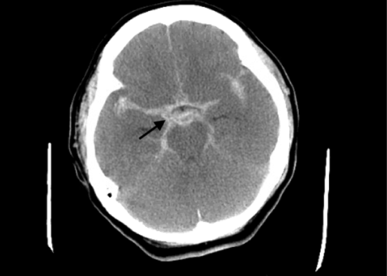
DOI: https://doi.org/10.21980/J8663NThe electrocardiogram demonstrated sinus tachycardia with ST segment elevation in lead aVR (black arrows) and diffuse ST depressions concerning for possible ST elevation myocardial infarction (STEMI). Given the events reported and the patient’s neurologic exam without sedation, non-contrast CT of the head was ordered; imaging showed evidence of a large subarachnoid hemorrhage, mostly at the level of the Circle of Willis (black arrow) concerning for an aneurysmal bleed as well as mild generalized white matter density suggestive of cerebral edema.
A Case Report of Epidural Hematoma After Traumatic Brain Injury
DOI: https://doi.org/10.21980/J8R059Non-contrast CT head demonstrated a right sided EDH (red arrow) with overlying scalp hematoma, left-sided subdural hematoma (blue arrow), and bilateral subarachnoid hemorrhages. No skull fractures were noted.
A Case Report on Miliary Tuberculosis in Acute Immune Reconstitution Inflammatory Syndrome
DOI: https://doi.org/10.21980/J81H02A portable single-view radiograph of the chest was obtained upon the patient’s arrival to the ED resuscitation bay that showed diffuse reticulonodular airspace opacities (red arrows) seen throughout the bilateral lungs, concerning for disseminated pulmonary tuberculosis. Subsequently, a computed tomography (CT) angiography of the chest was obtained which again demonstrates this diffuse reticulonodular airspace opacity pattern (red arrows).
Loose PEG Tube Leading to Peristomal Leakage and Peritonitis
DOI: https://doi.org/10.21980/J8HS7TFrontal chest X-ray showed a large radiolucent area (pink highlighted area) underneath the diaphragm (yellow line) and on top of the liver (blue highlighted area) and spleen (green highlighted area) suggestive of pneumoperitoneum possibly caused by gastrointestinal perforation. This large radiolucent area can also be seen underneath the diaphragm in the lateral view chest X-ray. Computed tomography (CT) was not performed due to his physical exam findings and the significant positive findings on chest X-ray. Surgery was consulted and patient was taken emergently to the operating room.
Rapid Airway Narrowing Associated with Hodgkin’s Lymphoma
DOI: https://doi.org/10.21980/J86D3QNeck X-ray showed nonspecific significant prevertebral soft tissue swelling at the level of the cervical spine, with associated apparent thickening of the epiglottis (yellow arrow), diffuse soft tissue swelling of the neck (red arrows) and tracheal airway narrowing (light blue arrow). The computed tomography imaging of the neck was significant for multiple conglomerating pathological lymph nodes with a significant mass effect (orange arrows) compressing the right internal jugular vein (green arrow).
Fitz Hugh Curtis Case Report
DOI: https://doi.org/10.21980/J82K9GA sagittal view from computed tomography (CT) of the abdomen and pelvis demonstrated fat stranding beneath the inferior margin of the liver (outlined in red). The axial view showed fat stranding adjacent to the ascending colon without significant colon wall thickening (arrow). Fat stranding can occur as a hazy increased attenuation (brightness) or a more distinct reticular pattern.


