Renal/Electrolytes
A Patient with Generalized Weakness – A Case Report
DOI: https://doi.org/10.21980/J8593CThe CT of the abdomen and pelvis showed evidence of a large subcapsular rim-enhancing fluid collection with multiple gas and air-fluid levels along the right kidney measuring 8 x 4 cm axially and 11 cm craniocaudally (blue outline) with mass effect on the right renal parenchyma (yellow outline). Another suspected fluid collection adjacent to the upper pole of the right kidney measuring 4 x 3.4 cm was noted (red outline). Bilateral pyelonephritis was suggested without hydronephrosis or nephrolithiasis. The findings suggested complicated pyelonephritis with emphysematous abscess and hematoma formation.
Case Report of a Pelvic Kidney with Ureteral Obstruction from Inguinal Hernia Entrapment and Concurrent Cryptorchid Testis
DOI: https://doi.org/10.21980/J8F345The patient was afebrile with normal lactate and white blood cell count. Initial CT imaging showed an ectopic right pelvic kidney with entrapment of his right ureter within an indirect right inguinal hernia causing severe hydronephrosis (coronal: white arrow). Also discovered was an ovoid hypodensity in the right anterior pelvis consistent with right undescended testis (axial: orange arrow; coronal: green arrow) that was previously unknown to the patient, with a normal left scrotal testis (axial: red arrowhead; coronal: blue arrowhead). Other potential etiologies of the patient’s symptoms could include appendicitis or incarcerated inguinal hernia, though the imaging results and absence of systemic inflammatory response syndrome made these causes less likely.
Case Report of Unusual Facial Swelling in an 8-Month-Old
DOI: https://doi.org/10.21980/J8M06FFacial ultrasound revealed local inflammatory changes such as increased echogenicity and heterogeneity in the soft tissues of the right cheek, suggestive of soft tissue edema. There was evidence of a prominent right parotid gland with increased heterogeneity suggestive of a traumatic injury. Additionally, facial ultrasound demonstrated a 6mm ill-defined anechoic collection within the right cheek without increased doppler flow (green arrow), thought to represent a focal area of edema instead of an abscess.
Case Report: Not Your Typical Kidney Stone
DOI: https://doi.org/10.21980/J8GD2TThe CT scan demonstrates nephrolithiasis with associated forniceal rupture. Encircled in the yellow outline is fluid, demonstrating a forniceal rupture. The stone is in the proximal aspect of the ureter, as highlighted by the purple arrow.
Bladder Diverticulum – A Case Report
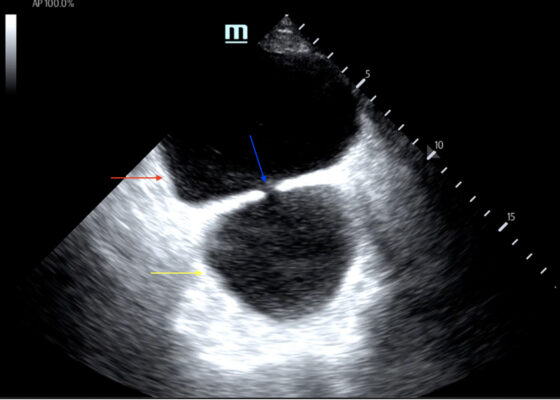
DOI: https://doi.org/10.21980/J8635COn examination, the patient was alert and oriented but in mild distress. Suprapubic fullness was noted upon abdominal palpation. Point of care ultrasound of the bladder showed two enlarged “bladders” with a central communication. Bedside total bladder volume was measured to be 1288 cm3 (the top “bladder” was measured to be 1011 cm3, while the bottom “diverticulum” was measured to be 277 cm3) by ultrasound.
The POCUS stills of the patient’s bladder demonstrated the bladder (red arrow) and bladder diverticulum (yellow arrow) with a central communication (blue arrow) in the transverse and sagittal views.
Hemorrhagic Renal Cyst
DOI: https://doi.org/10.21980/J8C92VBedside renal ultrasound demonstrated a right renal cyst with echogenic debris consistent with a hemorrhagic cyst (red arrow). In addition, a computed tomography (CT) scan of the abdomen and pelvis revealed a 4mm non-obstructing right renal stone and bilateral renal cysts. The CT also confirmed the ultrasound finding of a right renal cyst with mild perinephric stranding possibly consistent with a hemorrhagic cyst.
FAST Exam to Diagnose Subcapsular Renal Hematoma
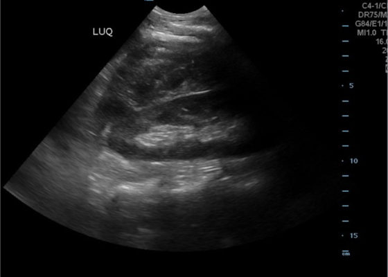
DOI: https://doi.org/10.21980/J8NP8DA bedside point of care ultrasound FAST exam was performed revealing a left subcapsular renal hematoma. The hematoma was a non-compressing hematoma, evidenced by preserved renal contour with the hematoma labeled with a red H and the normal renal contour labeled with a green K. Additionally, cortical necrosis and ischemia can be characterized by a dark, hypoechogenic renal cortex on ultrasonography with a decrease in flow to the cortex on color doppler which was not seen on this patient, providing further evidence that the hematoma was non-compressing. The hematoma was concluded to be an acute process due to its hypoechoic appearance with some mixed ultrasonographic echoes caused by the early deposit of fibrin.
Erectile Dysfunction as a Presenting Symptom for Renal Cell Carcinoma
DOI: https://doi.org/10.21980/J8563BThe MRI showed extensive spondylotic changes suggestive of malignancy (red arrows) with severe spinal canal stenosis at the lumbar spine L3-L4 (purple arrows) level contributing to clumping of cauda equina nerve roots and severe bilateral neuroforaminal narrowing with diffuse disc bulges abutting the exiting nerve roots at multiple levels. Findings also showed a hypo-attenuated tumor (blue arrow) and hyper-attenuated loculated tumor (green arrow) consistent with renal cell carcinoma (RCC).
A Woman with Arm Spasms
DOI: https://doi.org/10.21980/J8VP88The patient had a witnessed episode of isolated left upper extremity jerking, shown in the video, during which she was completely awake and conversant. Lab results were significant for serum glucose of 1167 mg/dL, no anion gap, and negative serum/urine ketones. She had a computed tomography (CT) of the head that did not show any acute pathology, and underwent a brain magnetic resonance imaging (MRI) without any signs of stroke or other pathology, shown below.
Computed Tomography and Ultrasound Diagnosis of Spontaneous Subcapsular Renal Hematoma
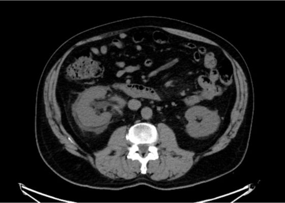
DOI: https://doi.org/10.21980/J8062DBedside ultrasound was performed and demonstrated a hypoechoic area within the left kidney (images not shown). The non-contrast computed tomography (CT) of the abdomen and pelvis shows a significantly enlarged left kidney and a region of high-attenuation encapsulating the left kidney, concerning for acute hemorrhage.
1›
Page 1 of 2


