Acute Pancreatitis
History of present illness:
A 28-year-old female presented to the emergency department with slow onset epigastric abdominal pain that radiated to her back for one day. The patient described the pain as sharp, constant, and 9/10 in severity. Her pain was associated with non-bilious, non-bloody vomiting. On physical exam, the patient had tenderness in the epigastric region. Pregnancy test was negative. There was no evidence of periumbilical or flank ecchymosis. Due to the severity of her pain, a computed tomography (CT) scan of her abdomen and pelvis was ordered.
Significant findings:
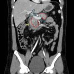
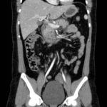
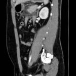
Computed tomography of the abdomen and pelvis with contrast show edema of the pancreas (red outline) and duodenum (yellow arrow) with peripancreatic inflammation, fluid and fat stranding (blue highlight). The distal pancreatic tail was noted to appear normal (green arrow). There was no organized drainable fluid collection, and no parenchymal hypo-enhancement. These findings are consistent with moderate severity acute interstitial pancreatitis.
Discussion:
Pancreatitis is one of the most common diagnoses in hospitalizations among gastrointestinal disorders, accounting for more than 200,000 admissions each year.1-3 Premature activation of trypsin in the acinar cells of the pancreas leads to activation of proteolytic enzymes. Subsequent leakage of fluid into the pancreas and surrounding tissues causing an inflammatory response.3-5 Although most cases of pancreatitis are mild in nature, 10%-20%of cases lead to systemic inflammatory response syndrome (SIRS), which can predispose patients to multiple organ system failure and pancreatic necrosis.4,6 Common complications of acute pancreatitis include pancreatic fluid collections and the development of pseudocysts, which may require surgical intervention.6 The most common risk factors for pancreatitis are cholelithiasis and alcoholism.7Clinical features include epigastric abdominal pain radiating to the back, nausea, and vomiting. Although serum lipase is often found to be elevated in acute pancreatitis, a normal serum lipase does not exclude the diagnosis of acute pancreatitis.2,6 The diagnosis of acute pancreatitis typically requires two of the three following findings: abdominal pain characteristic of acute pancreatitis, serum amylase or lipase greater than three times the upper limit of normal and/or findings of acute pancreatitis on CT.4
Although imaging is not required for the diagnosis, and the majority of patients will likely not receive imaging, information obtained from radiologic studies may elucidate possible etiologies, severity, and sequelae of acute pancreatic necrosis (APN).5 Computed tomography is the most common imaging modality for suspected acute pancreatitis with a sensitivity and specificity of 78% and 86% respectively.7 Common findings include focal or diffuse enlargement and heterogenous enhancement of the pancreas.8 However, magnetic resonance imaging (MRI) is better able to characterize fluid collections and to detect early pancreatitis.5,6 The patient in this case had a normal lipase and pain consistent with acute pancreatitis; because it was a first episode and she had significant abdominal pain, she received a CT of her abdomen and pelvis to evaluate for possible etiologies or complications.
Upon diagnosis, admission for pain control and aggressive intravenous fluid replacement is often needed for symptomatic control and is important in avoiding development of necrotizing pancreatitis.4 Pancreatic necrosis can also develop within the first few days and can lead to hypovolemia, hypotension, systemic inflammatory response syndrome, or even death.4,6 Prophylactic antibiotics are not recommended. However, if there is concern for infection, broad spectrum coverage, including coverage for gram negative and anaerobic organisms should be utilized. Approximately 20% of patients with APN develop extrapancreatic infections.9 Further stratification to a monitored versus critical-care setting should be made judiciously using the patient’s clinical status and comorbidities. Risk Calculators, such as APACHE II, may also be used to help appropriately make dispositions for the patient.
This patient was admitted for pain control and fluid hydration; she improved and was discharged a few days later. Unfortunately, the etiology of the patient’s pancreatitis was not evident at the time of discharge.
Topics:
Acute pancreatitis, fluid collection, gastrointestinal.
References:
- DeFrances CJ, Hall MJ, Podgornik MN. 2003 National Hospital Discharge Survey. Advance data from vital and health statistics. No. 359. Hyattsville, Md.: National Center for Health Statistics. 2005.
- Dhaka N, Samanta J, Kochhar S, et al. Pancreatic fluid collections: what is the ideal imaging technique? World J Gastroenterol. 2015;21(48):13403-10. doi: 10.3748/wjg.v21.i48.13403
- Peery AF, Dellon ES, Lund J, et al. Burden of gastrointestinal disease in the United States: 2012 update. Gastroenterology. 2012;143(5):1179-1187.e1-3. doi: 10.1053/j.gastro.2012.08.002
- Busireddy KK, AlObaidy M, Ramalho M, et al. Pancreatitis-imaging approach. World J Gastrointest Pathophysiol. 2014;5(3):252-270. doi: 10.4291/wjgp.v5.i3.252
- Whitcomb DC. Clinical Practice. Acute pancreatitis. N Engl J Med. 2006;354(20):2142-2150.doi:10.1056/NEJMcp054958
- Banks PA, Freeman ML. Practice Parameters Committee of the American College of Gastroenterology. Practice guidelines in acute pancreatitis. Am J Gastroenterol. 2006;101(10):2379-2400. doi: 10.1111/j.1572-0241.2006.00856.x
- Carroll JK, Herrick B, Gipson T, Lee SP. Acute pancreatitis: diagnosis, prognosis, treatment. Am Fam Physician. 2007;75(10):1513-1520.
- Banks PA, Bollen TL, Dervenis C, et al. Classification of acute pancreatitis–2012: revision of the Atlanta classification and definitions by international consensus. Gut. 2013;62(1):102–111. doi: 10.1136/gutjnl-2012-302779
- Besselink MG, van Santvoort HC, Boermeester MA, et al. Timing and impact of infections in acute pancreatitis. Br J Surg. 2009;96(3):267-273. doi: 10.1002/bjs.6447


