• Registration
  • Login
JETem
  • Home
  • About
    • Aim and Scope
    • Our Team
    • Editorial Board
    • FAQ
  • Issues
    • Current Issue
    • Ahead of Print
    • Past Issues
  • Visual EM
    • Latest Visual EM
    • Search Visual EM
    • Thumbnail Library
  • For Authors
    • Instructions for Authors
    • Submit to JETem
    • Photo Consent
    • Policies
      • Peer Review Policy
      • Copyright Policy
      • Editorial Policy, Ethics and Responsibilities
      • Conflicts of Interest & Informed Consent
      • Open Access Policy
  • For Reviewers
    • Instructions for JETem Reviewers
    • Interested in Being a JETem Reviewer?
  • Topic
    • Abdominal / Gastroenterology
    • Administration
    • Board Review
    • Cardiology / Vascular
    • Clinical Informatics, Telehealth and Technology
    • Dermatology
    • EMS
    • Endocrine
    • ENT
    • Faculty Development
    • Genitourinary
    • Geriatrics
    • Hematology / Oncology
    • Infectious Disease
    • Miscellaneous
    • Neurology
    • Ob / Gyn
    • Ophthalmology
    • Orthopedics
    • Pediatrics
    • Pharmacology
    • Procedures
    • Psychiatry
    • Renal / Electrolytes
    • Respiratory
    • Social Determinants of Health
    • Toxicology
    • Trauma
    • Ultrasound
    • Urology
    • Wellness
    • Wilderness
  • Modality
    • Curricula
    • Innovations
    • Lectures
    • Certifying Exam Practice Cases
      • Clinical Care Cases
      • Communication & Procedural Cases
      • Traditional Oral Boards
    • Podcasts
    • Simulation
    • Small Group Learning
    • Team Based Learning
    • Visual EM
  • Contact Us

X-Ray

Creative Commons images

Empyema

Jonathan Patane, MD* and Daniel Ritter, BS^

DOI: https://doi.org/10.21980/J86P9RIssue 3:1 No ratings yet.
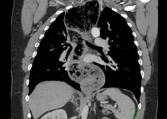
The chest X-ray shows a large fluid collection in the right lung demonstrated by the opacification that blunts the costophrenic angle on the right side. There is also a meniscus present, which is generally indicative of fluid.  Chest computed tomography (CT) demonstrated an infiltrate with a mixture of densities within the same collection, consistent with a loculated effusion and concerning for an empyema.
RespiratoryInfectious DiseaseVisual EM
Creative Commons images

Bilateral Hip Dislocation in Unrestrained Driver

Samer Assaf, MD* and Ghadi Ghanem*

DOI: https://doi.org/10.21980/J8HD0C Issue 2:4 No ratings yet.
The initial radiograph of the pelvis revealed bilateral hip dislocations. Small bony fragments were noted in the right hip joint, suggestive of an underlying fracture. The sacroiliac joints and the pelvic ring were intact. In the emergency department, bilateral hip reductions were performed using the Captain Morgan technique.1 The post-reduction film showed reduction of the bilateral hip dislocations with extensive comminuted and displaced fractures of the right and left acetabula.
OrthopedicsVisual EM
Creative Commons images

Open Book Pelvic Fracture

Samer Assaf, MD* and Ghadi Ghanem*

DOI: https://doi.org/10.21980/J8CK7H Issue 2:3 No ratings yet.
The initial radiograph of the pelvis shows an open-book pelvic fracture deformity with pubic symphyseal dislocation, left greater than right sacroiliac diastases, and fractures of the left superior and inferior pubic rami, right inferior pubic ramus, and left acetabular anterior column. The additional inlet and outlet radiographs of the pelvis after application of a pelvic binder also show an open book fracture with significant improvement of the widened pubic symphysis.
OrthopedicsTraumaVisual EM
Creative Commons images

Spontaneous Pneumothorax

John Costumbrado, MD, MPH* and Steven Kim, MD*

DOI: https://doi.org/10.21980/J8M33BIssue 2:4 No ratings yet.
Initial chest radiograph showed a 50% right-sided pneumothorax with no mediastinal shift, which can be identified by the sharp line representing the pleural lung edge (see arrows) and lack of peripheral lung markings extending to the chest wall. While difficult to accurately estimate volume from a two-dimensional image, a 2 cm pneumothorax seen on chest radiograph correlates to approximately 50% volume.1 The patient underwent insertion of a pigtail pleural drain on the right and repeat chest radiograph showed resolution of previously seen pneumothorax. Ultimately the pigtail drain was removed and chest radiograph showed clear lung fields without evidence of residual pneumothorax or pleural effusion.
RespiratoryVisual EM
Creative Commons images

Pediatric Esophageal Foreign Body

Samer Assaf, MD* and Ryan Gibney, BS*

DOI: https://doi.org/10.21980/J8GD1F Issue 2:4 No ratings yet.
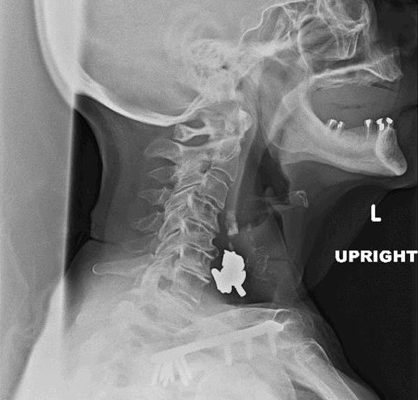
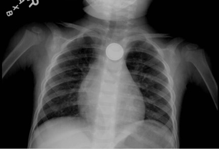
A radiopaque foreign body was visualized in the proximal esophagus at the thoracic inlet on the chest and neck radiographs. The foreign body appeared to be metallic with visualized concentric rings consistent with a coin.
Abdominal/GastroenterologyPediatricsVisual EM
Creative Commons images

Acetabular Fracture

Chad Correa* and Sari Lahham, MD^

DOI: https://doi.org/10.21980/J8BK8K Issue 2:4 No ratings yet.
The non-contrast CT images show a minimally displaced comminuted fracture of the right acetabulum involving the acetabular roof, medial and anterior walls (red arrows), with associated obturator muscle hematoma (blue oval).
OrthopedicsVisual EM
Creative Commons images

Volvulus

Sari Lahham, MD, MBA*, Kathryn Bennett, BS* and Mohammad Helmy, MD†

DOI: https://doi.org/10.21980/J8JH0Q Issue 2:3 No ratings yet.
Upright and supine frontal radiographs of the abdomen demonstrate gas dilation of the large bowel from the level of the cecum to the sigmoid colon with air fluid levels (yellow arrows). There is a swirled configuration of the distal descending to sigmoid colon indicating the level of the volvulus (dashed yellow line) and giving rise to the classic “coffee bean” sign (dotted white tracing). Note the elevated left hemidiaphragm on the upright view reflecting abdominal distention with increased intra-abdominal pressure (red arrow).
Abdominal/GastroenterologyVisual EM
Creative Commons images

Re-expansion Pulmonary Edema

Kathryn Bennett, BS, BA* and Benjamin Feldman, MD*

DOI: https://doi.org/10.21980/J8WS6V Issue 2:3 No ratings yet.
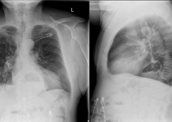
Initial chest X-ray (chest X-ray 1) showed a right-sided pleural effusion with compressive atelectasis of the mid to lower right lung. Repeat chest X-ray immediately after evacuation (chest X-ray 2) shows improvement of the pleural effusion and a new trace apical right pneumothorax measuring 6.7 mm. When the patient became tachypneic, a third X-ray (chest X-ray 3) showed persistent trace apical right pneumothorax measuring 6.7 mm.
RespiratoryVisual EM
Creative Commons images

Open Pneumothorax

Bart Paull, MD*, Zachary Jarou, MD* and David Richards, MD*

DOI: https://doi.org/10.21980/J88036 Issue 2:3 No ratings yet.
A large chest wound was clinically obvious. A chest radiograph performed after intubation showed subcutaneous emphysema, an anterior rib fracture, and a right-sided pneumothorax. He was then taken to the operating room for further management.
TraumaRespiratoryVisual EM
Creative Commons images

Supracondylar Fracture

Jessica Andrusaitis, BS, MS* and Ben Feldman, MD*

DOI: https://doi.org/10.21980/J8492P Issue 2:3 No ratings yet.
History of present illness: A 15-year-old male presented to the emergency department with right elbow pain after falling off a skateboard.  The patient denied a decrease in strength or sensation but did endorse paresthesias to his hand.  On exam, the patient had an obvious deformity of his right elbow with tenderness to palpation and decreased range of motion at the
OrthopedicsVisual EM
«‹7›»
Page 7 of 10

JETem is an online, open access, peer-reviewed, journal-repository for EM educators. We are PMC Indexed.

Most Viewed

  • A Longitudinal, Practical Curriculum for Faculty Development as New Coaches in Graduate Medical Education
  • Cognitive Errors and Debiasing
  • Critical Care Transport: Blunt Polytrauma in Pregnancy
  • Beta-Blocker Toxicity
  • A Comprehensive and Modality Diverse Cervical Spine and Back Musculoskeletal Physical Exam Curriculum for Medical Students

Visit Our Collaborators

Creative Commons Licence
This work is licensed under a Creative Commons Attribution 4.0 International License.

About

Education

Learners should benefit from active learning. JETem accepts submissions of team-based learning, small group learning, simulation, podcasts, lectures, innovations, curricula, question sets, and visualEM.

Scholarship

We believe educators should advance through the scholarship of their educational work. JETem gives educators the opportunity to publish scholarly academic work so that it may be widely distributed, thereby increasing the significance of their results.

Links

  • Home
  • Aim and Scope
  • Current Issue
  • For Reviewers
  • Instructions for Authors
  • Contact Us

Newsletter

Sign up to receive updates from JETem regarding newly published issues and findings.

Copyright Creative Commons Attribution 4.0 International