
X-Ray
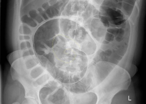

Volvulus
DOI: https://doi.org/10.21980/J8JH0QUpright and supine frontal radiographs of the abdomen demonstrate gas dilation of the large bowel from the level of the cecum to the sigmoid colon with air fluid levels (yellow arrows). There is a swirled configuration of the distal descending to sigmoid colon indicating the level of the volvulus (dashed yellow line) and giving rise to the classic “coffee bean” sign (dotted white tracing). Note the elevated left hemidiaphragm on the upright view reflecting abdominal distention with increased intra-abdominal pressure (red arrow).

Re-expansion Pulmonary Edema
DOI: https://doi.org/10.21980/J8WS6VInitial chest X-ray (chest X-ray 1) showed a right-sided pleural effusion with compressive atelectasis of the mid to lower right lung. Repeat chest X-ray immediately after evacuation (chest X-ray 2) shows improvement of the pleural effusion and a new trace apical right pneumothorax measuring 6.7 mm. When the patient became tachypneic, a third X-ray (chest X-ray 3) showed persistent trace apical right pneumothorax measuring 6.7 mm.

Open Pneumothorax
DOI: https://doi.org/10.21980/J88036A large chest wound was clinically obvious. A chest radiograph performed after intubation showed subcutaneous emphysema, an anterior rib fracture, and a right-sided pneumothorax. He was then taken to the operating room for further management.

Supracondylar Fracture
DOI: https://doi.org/10.21980/J8492PHistory of present illness: A 15-year-old male presented to the emergency department with right elbow pain after falling off a skateboard. The patient denied a decrease in strength or sensation but did endorse paresthesias to his hand. On exam, the patient had an obvious deformity of his right elbow with tenderness to palpation and decreased range of motion at the
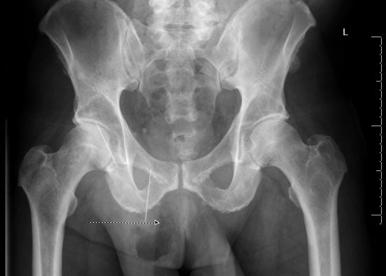
Bowel Perforation complicating an incarcerated inguinal hernia
DOI: https://doi.org/10.21980/J8D30BThe AP and lateral pelvis x-rays revealed two sewing needles, 60 mm in length, within the soft tissue over the anterior right lower hemipelvis. In addition, the AP view showed emphysema involving the right hemiscrotum (arrow), concerning for perforated bowel.

An Elderly Female with Dyspnea and Abdominal Pain
DOI: https://doi.org/10.21980/J83S3KRadiography shows a dilated, gas-filled structure that fills nearly the entire left hemi-thorax. Lung markings are visible in the uppermost portion of the left hemi-thorax. There is mediastinal shift to the right. In the visualized portion of the abdomen, dilated loops of bowel are also visualized. This constellation of findings is consistent with a tension gastrothorax.
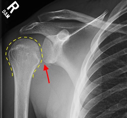
Irreducible Traumatic Posterior Shoulder Dislocation
DOI: https://doi.org/10.21980/J8V884Radiographs demonstrated posterior displacement of the humeral head on the “Y” view (see white arrow) and widening of the glenohumeral joint space on anterior-posterior view (see red arrow). The findings were consistent with posterior dislocation and a Hill-Sachs type deformity. Sedation was performed and reduction was attempted using external rotation, traction counter-traction. An immediate “pop” was felt during the procedure. Post-procedure radiographs revealed a persistent posterior subluxation with interlocking at posterior glenoid. CT revealed posterior dislocation with acute depressed impaction deformity medial to the biceps groove with the humeral head perched on the posterior glenoid, interlocked at reverse Hill-Sachs deformity (see blue arrow).
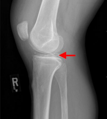
Pseudogout and Calcium Pyrophosphate Disease
DOI: https://doi.org/10.21980/J8QG66Radiographs of the knee showed multiple radio-dense lines paralleling the articular surface (see red arrows) consistent with calcium pyrophosphate crystal deposition within the joint often seen in calcium pyrophosphate disease (CPPD) also known as pseudogout.