Pediatric Esophageal Foreign Body
History of present illness:
A 3-year-old boy was brought in to the emergency department by his parents after suspected foreign body ingestion 45 minutes prior to arrival. The patient’s mother stated that he likely swallowed an arcade token. The patient was noted to have a brief choking episode at home followed by persistent drooling and vomiting. Parents denied difficulty breathing or abdominal pain. On exam, the patient had normal vitals, yet appeared uncomfortable with active drooling. However, his airway was patent without stridor, and his oropharyngeal and abdominal exams were unremarkable.
Significant findings:
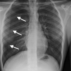
A radiopaque foreign body was visualized in the proximal esophagus at the thoracic inlet on the chest and neck radiographs. The foreign body appeared to be metallic with visualized concentric rings consistent with a coin.
Discussion:
Esophageal foreign bodies (FB) contribute to pediatric morbidity and mortality in the United States. Of the common items ingested, coins are the most common at 88%.1 Button batteries, which can lead to severe esophageal injury, have become more common with prevalence of small electronics in households.2 Small foreign bodies can be managed expectantly if they progress into gastrointestinal tract; however pediatric patients often present with objects lodged at the level of C6 due to the physiologic narrowing at the cricopharyngeal muscle.3
Complications of symptomatic pediatric FB ingestions such as esophageal necrosis, perforation, agitation, and airway compromise are particularly dangerous and warrant rapid diagnosis and intervention.4-5 Current guidelines recommend flexible endoscopy under anesthesia with endotracheal intubation for airway protection. There are several other potential modalities for management that may be successful and less invasive. Placement of Foley catheter distal to foreign body, inflation, and traction with extraction is a fast, safe, and cost-effective procedure that can be done bedside under limited procedural anesthesia.3-6
Our patient underwent rapid sequence intubation in the emergency department and had successful extraction of the coin under direct visualization using Magill forceps, followed by extubation and complete resolution of all symptoms.
Topics:
Esophageal foreign body, X-ray, radiograph, pediatric foreign body, coin.
References:
- Little DC, Shah SR, St Peter SD, Calkins CM, Morrow SE, Murphy JP, et al. Esophageal foreign bodies in the pediatric population: our first 500 cases. J Pediatr Surg. 2006;41:914-918. doi: 10.1016/j.jpedsurg.2006.01.022
- Jatana KR, Rhoades K, Milkovich S, Jacobs IN. Basic mechanism of button battery ingestion injuries and novel mitigation strategies after diagnosis and removal. Laryngoscope. 2017(6);1276-1282. doi: 10.1002/lary.26362
- Uyemura MC. Foreign body ingestion in children. Am Fam Physician. 2005;72(2):287.
- Patoulias D, Patoulias I, Kaselas C, Feidantsis T, Farmakis K, Kalogirou M. Lower esophageal sphincter relaxation by administrating hyoscine-N-butylbromide for esophageal impaction by coin-shaped foreign bodies; prospective clinical study in pediatric population. Folia Med Cracov. 2016;56(4):21-29.
- Waltzman ML. Management of esophageal coins. Curr Opin Pediatr. 2006;18(5):571. doi: 10.1097/01.mop.0000245361.91077.b5
- Burgos A, Rábago L, Triana P. Western view of the management of gastroesophageal foreign bodies. World J Gastrointest Endosc. 2016;8(9),378-384. doi: 10.4253/wjge.v8.i9.378


