
X-Ray


Gastric Volvulus
DOI: https://doi.org/10.21980/J8335FPoint of care ultrasound of his abdomen showed a large fluid filled structure with well-defined borders containing gastric contents extending from the xiphoid process to the umbilical region. No free fluid was noted on focus assessment with sonography for trauma (FAST) examination. A computed tomography (CT) scan was performed emergently and it was noted that the patient had a significantly distended stomach and gastric volvulus (blue arrows) noted in the area of his paraesophageal/hiatal hernia.

Diagnosis and Treatment of an Anterior Shoulder Dislocation with Bedside Ultrasound
DOI: https://doi.org/10.21980/J8Z924Bedside ultrasound with the transducer placed on the posterior right shoulder revealed an anterior dislocation of the right humerus. This is evident by displacement of the humeral head further away from the posteriorly placed ultrasound transducer, and appears deep to the glenoid cavity. In a posterior shoulder dislocation, the humeral head would appear closer to the transducer (and the near field of the ultrasound image) than the glenoid. Note that a hypoechoic, heterogeneous fluid collection is within the joint space, compatible with a hematoma. A right shoulder X-ray confirmed the anterior dislocation with no evidence of fracture. Under direct ultrasound guidance the glenohumeral joint space was injected with 10 mL of 2% lidocaine as an intraarticular anesthetic block. The right shoulder was reduced using continual traction. Post-reduction ultrasound demonstrated a successful shoulder reduction, depicted by the humeral head being relocated to its anatomical location, adjacent to the glenoid cavity, as noted on the ultrasound image. A hematoma remains present within the joint space. Successful shoulder reduction was further confirmed by X-ray. The patient’s arm was placed in a sling and she was discharged home with orthopedics follow-up.
Pneumocystis jirovecii (carinii) Pneumonia
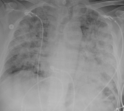
DOI: https://doi.org/10.21980/J8RW6NChest X-ray showed diffuse, patchy interstitial and alveolar infiltrates bilaterally concerning for Pneumocystis jirovecii(previously Pneumocystis carinii) pneumonia (PJP). The AP radiograph (top left figure) showed the classic “bat-wing” distribution on the left side. Repeat radiograph (bottom figure) one day after admission showed worsening of the infiltrates.

Bilateral Shoulder Dislocation after Ski Injury
DOI: https://doi.org/10.21980/J86929An anteroposterior chest X-ray demonstrates bilateral shoulder dislocations. Both the right and left humeral heads (blue lines) are displaced medially, anteriorly, and inferiorly from their normal positions in the glenoid fossae (red lines), thus signifying bilateral anterior dislocations. There is also a fracture of the left humeral head at the greater tubercle (green arrow).
Talonavicular Dislocation
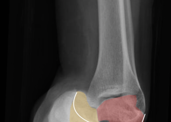
DOI: https://doi.org/10.21980/J8PG91The X-rays were significant for a subtalar dislocation. The calcaneus (red) is laterally displaced with respect to the talar head (orange), and the white lines indicate the normal articular surface. Additionally, there was a talonavicular dislocation, as seen in the fourth image: the talus (green) and navicular bone (purple) overlapping suggests a dislocation. In a normally aligned foot, the boundaries of the two bones create a point of articulation.
Endocarditis
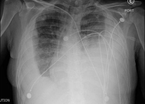
DOI: https://doi.org/10.21980/J8JP73Upright frontal radiograph of the chest demonstrated large pleural effusion on the left and moderate pleural effusion on the right as shown by the visible menisci on both sides (red arrows) with diffuse bilateral nodular densities (yellow dotted lines), consistent with septic pulmonary emboli. Computed tomography (CT) of the chest demonstrated multiple scattered lung nodules bilaterally containing internal foci of air cavitation (green dotted lines).

Subcutaneous Emphysema After Chest Trauma
DOI: https://doi.org/10.21980/J8864NPlain film anteroposterior (AP) radiography of the chest shows left-sided subcutaneous emphysema (red arrow) with overlapping muscle striations of the pectoralis major (green arrow). After chest tube placement (blue arrow), AP chest radiography shows persistent left-sided subcutaneous emphysema (red arrow). CT of the chest shows pneumomediastinum (blue arrow), left apical pneumothorax (pink arrow), and subcutaneous emphysema (red arrow) at the level of T2. At the level of T6, rib fractures can be visualized on the CT (yellow arrow). At the level of T8, left sided pneumothorax is also seen (pink arrow) as the absence of lung tissue on CT.
An Unusual Case of Hematemesis
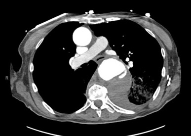
DOI: https://doi.org/10.21980/J84H00The patient’schest X-ray revealed a prominent mediastinum and opacification in the left middle and lower lung fields. The CT showed an aortic aneurysm extending from the thorax to the abdomen with rupture near T7 (blue arrow). It also showed periaortic hemorrhage with active extravasation (green arrow) likely secondary to a penetrating ulcer and bilateral pulmonary opacities concerning for hemothorax (pink arrow).