• Registration
  • Login
JETem
  • Home
  • About
    • Aim and Scope
    • Our Team
    • Editorial Board
    • FAQ
  • Issues
    • Current Issue
    • Ahead of Print
    • Past Issues
  • Visual EM
    • Latest Visual EM
    • Search Visual EM
    • Thumbnail Library
  • For Authors
    • Instructions for Authors
    • Submit to JETem
    • Photo Consent
    • Policies
      • Peer Review Policy
      • Copyright Policy
      • Editorial Policy, Ethics and Responsibilities
      • Conflicts of Interest & Informed Consent
      • Open Access Policy
  • For Reviewers
    • Instructions for JETem Reviewers
    • Interested in Being a JETem Reviewer?
  • Topic
    • Abdominal / Gastroenterology
    • Administration
    • Board Review
    • Cardiology / Vascular
    • Clinical Informatics, Telehealth and Technology
    • Dermatology
    • EMS
    • Endocrine
    • ENT
    • Faculty Development
    • Genitourinary
    • Geriatrics
    • Hematology / Oncology
    • Infectious Disease
    • Miscellaneous
    • Neurology
    • Ob / Gyn
    • Ophthalmology
    • Orthopedics
    • Pediatrics
    • Pharmacology
    • Procedures
    • Psychiatry
    • Renal / Electrolytes
    • Respiratory
    • Social Determinants of Health
    • Toxicology
    • Trauma
    • Ultrasound
    • Urology
    • Wellness
    • Wilderness
  • Modality
    • Curricula
    • Innovations
    • Lectures
    • Certifying Exam Practice Cases
      • Clinical Care Cases
      • Communication & Procedural Cases
      • Traditional Oral Boards
    • Podcasts
    • Simulation
    • Small Group Learning
    • Team Based Learning
    • Visual EM
  • Contact Us

X-Ray

Creative Commons images

Subcutaneous Emphysema After Chest Trauma

Ronald Goubert, BS* and Alisa Wray, MD, MAEd*

DOI: https://doi.org/10.21980/J8864NIssue 3:4 No ratings yet.
Plain film anteroposterior (AP) radiography of the chest shows left-sided subcutaneous emphysema (red arrow) with overlapping muscle striations of the pectoralis major (green arrow). After chest tube placement (blue arrow), AP chest radiography shows persistent left-sided subcutaneous emphysema (red arrow). CT of the chest shows pneumomediastinum (blue arrow), left apical pneumothorax (pink arrow), and subcutaneous emphysema (red arrow) at the level of T2. At the level of T6, rib fractures can be visualized on the CT (yellow arrow). At the level of T8, left sided pneumothorax is also seen (pink arrow) as the absence of lung tissue on CT.
TraumaRespiratoryVisual EM
Creative Commons images

An Unusual Case of Hematemesis

Amanda Amen, BA*, Jane Xiao, MD^, Julie Parks-Bortel, MD† and Shanna Jones, MD†

DOI: https://doi.org/10.21980/J84H00Issue 3:4 No ratings yet.
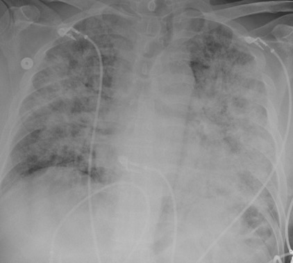
The patient’schest X-ray revealed a prominent mediastinum and opacification in the left middle and lower lung fields. The CT showed an aortic aneurysm extending from the thorax to the abdomen with rupture near T7 (blue arrow). It also showed periaortic hemorrhage with active extravasation (green arrow) likely secondary to a penetrating ulcer and bilateral pulmonary opacities concerning for hemothorax (pink arrow).
Cardiology/VascularVisual EM
Creative Commons images

Croup

Victor Cisneros, MD, MPH*, Joseph Zakaria, BS* and Wirachin Hoonpongsimanont, MD*

DOI: https://doi.org/10.21980/J8W05JIssue 3:4 No ratings yet.
The anteroposterior X-ray reveals the classic steeple sign (blue outline) indicative of subglottic edema leading to tracheal narrowing, consistent with croup. The lateral x-ray shows narrowing of the subglottic region (green outline and arrows).
PediatricsRespiratoryVisual EM
Creative Commons images

Don’t Forget the Pacemaker – A Rare Complication

Amanda Esposito, MD*, Kenneth Rapp, MD* and Albert Ritter, MD*

DOI: https://doi.org/10.21980/J8GS7HIssue 3:4 No ratings yet.
The ECG demonstrated the presence of pacemaker spikes without appropriate capture (green arrows) and a ventricular escape rhythm which can be identified by an absence of P waves prior to the QRS complex (purple arrows). The portable chest X- demonstrated displaced pacemaker leads (red arrows) that were coiled around the pulse generator (blue arrow).   
Cardiology/VascularVisual EM
Creative Commons images

The Role of Chest X-Ray and Bedside Ultrasound in Diagnosing Pulmonary Bleb versus Pneumothorax

Mohamad Moussa, MD* and Venkat Reddy Vaddamani, BS*

DOI: https://doi.org/10.21980/J8MP7QIssue 3:3 No ratings yet.
The patient was evaluated with bedside ultrasound for concern of possible pneumothorax. Imaging of the left lung with M-mode demonstrated a “sea shore” sign showing a wavy pattern below the pleural line caused by lung sliding as well as “comet tail” artifact caused by from the deep pleura. However, there was no lung sliding on the right shown by a lack of “comet tail” artifact and a “bar code” sign where M-mode shows straight lines throughout the image, this is caused by lack of motion below the pleura. This lack of lung sliding is consistent with possible pneumothorax or bleb.  A two-view chest X-ray (CXR) revealed absent lung parenchyma in the right lung similar to a large pneumothorax (see red outline). Electronic medical record chart review revealed previous CXRs with similar findings. This patient was determined to have an acute COPD exacerbation with chronic blebs, but no pneumothorax.
RespiratoryVisual EM
Creative Commons images

Bilateral Tibia/Fibula Fractures in Automobile versus Pedestrian Accident

Kevin Roy, BS* and Jonathan Peña, MD*

DOI: https://doi.org/10.21980/J8C636 Issue 3:3 No ratings yet.
Plain film shows severely comminuted and displaced mid tibia/fibula fractures of bilateral lower extremities (red arrows) and comminuted right fibular head (blue arrow) and proximal shaft fracture (yellow arrow).
OrthopedicsVisual EM
Creative Commons images

Dorsally-Displaced Metacarpal Dislocation-Fracture

Richard Barnett, DO* and Richard Brodsky, MD*

DOI: https://doi.org/10.21980/J8ZW54Issue 3:3 No ratings yet.
A two-view radiograph of the right hand was obtained which revealed a dorsal dislocation of the distal fourth and fifth metacarpals (see red and blue outline, respectively) with a concomitant fracture of the distal fifth metacarpal (see yellow line) and avulsion fracture of the lateral aspect of the hamate (see green line). After reduction the fourth and fifth metacarpal dislocations are resolved; however, the distal fifth metacarpal fracture (yellow line) and avulsion fracture of the lateral aspect of the hamate (green line) are still visible.
OrthopedicsVisual EM
Creative Commons images

Woman Swallows a “Handful of Pills”

Sarah E Mott, MD*, Michael Paddock, DO, MS*^ and Jessie Nelson, MD*^

DOI: https://doi.org/10.21980/J8V64XIssue 3:3 No ratings yet.
Soft tissue lateral X-ray of neck was performed. The lateral soft tissue X-ray of the neck showed a metallic foreign body at the level cricoid.
Abdominal/GastroenterologyVisual EM
Creative Commons images

Lisfranc Injury

Lisa Schwartz, BS*, Nelly Tan, MD^ and John Costumbrado, MD, MPH*

DOI: https://doi.org/10.21980/J8QD1MIssue 3:3 No ratings yet.
The frontal view of the right foot showed divergent dislocation of the second through fifth metatarsal bones (red outlines) consistent with Lisfranc injury. Though the Lisfranc ligament is not visualized by radiograph, the yellow markings represent the location of the Lisfranc ligament between the medial cuneiform (blue dot) and the base of the second metatarsal bone.  The first metatarsal and the medial cuneiform remain congruent. The lateral view shows dorsal dislocation of the midfoot (pink circle) consistent with instability. There is associated extensive midfoot soft tissue swelling.
OrthopedicsVisual EM
Creative Commons images

Incidental Hiatal Hernia on Chest X-ray

Ahmed Farhat, BS* and Daryn Towle, MD*

DOI: https://doi.org/10.21980/J8KP8SIssue 3:3 No ratings yet.
The two-view chest X-ray shows mild opacification of the bilateral lower lobes concerning for pneumonia (red arrows). Incidental retrocardiac opacity with air-fluid level consistent with large hiatal hernia is also observed (green arrow).
Abdominal/GastroenterologyVisual EM
«‹5›»
Page 5 of 10

JETem is an online, open access, peer-reviewed, journal-repository for EM educators. We are PMC Indexed.

Most Viewed

  • A Longitudinal, Practical Curriculum for Faculty Development as New Coaches in Graduate Medical Education
  • Cognitive Errors and Debiasing
  • Critical Care Transport: Blunt Polytrauma in Pregnancy
  • Beta-Blocker Toxicity
  • A Comprehensive and Modality Diverse Cervical Spine and Back Musculoskeletal Physical Exam Curriculum for Medical Students

Visit Our Collaborators

Creative Commons Licence
This work is licensed under a Creative Commons Attribution 4.0 International License.

About

Education

Learners should benefit from active learning. JETem accepts submissions of team-based learning, small group learning, simulation, podcasts, lectures, innovations, curricula, question sets, and visualEM.

Scholarship

We believe educators should advance through the scholarship of their educational work. JETem gives educators the opportunity to publish scholarly academic work so that it may be widely distributed, thereby increasing the significance of their results.

Links

  • Home
  • Aim and Scope
  • Current Issue
  • For Reviewers
  • Instructions for Authors
  • Contact Us

Newsletter

Sign up to receive updates from JETem regarding newly published issues and findings.

Copyright Creative Commons Attribution 4.0 International