Lisfranc Injury
History of present illness:
A 21-year-old male was brought in by ambulance to the emergency department status post motor vehicle accident. The patient was the restrained driver of a vehicle that struck a wall head-on at an unknown speed. His vitals were stable. The patient’s only complaint was severe pain to his right foot. On exam, the patient’s right foot was swollen and tender with deformity and was neurovascularly intact. Due to the unstable nature of the traumatic injury, the patient underwent urgent midfoot stabilization in the operating room.
Significant findings:
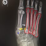
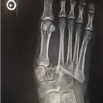
The frontal view of the right foot showed divergent dislocation of the second through fifth metatarsal bones (red outlines) consistent with Lisfranc injury. Though the Lisfranc ligament is not visualized by radiograph, the yellow markings represent the location of the Lisfranc ligament between the medial cuneiform (blue dot) and the base of the second metatarsal bone. The first metatarsal and the medial cuneiform remain congruent. The lateral view shows dorsal dislocation of the midfoot (pink circle) consistent with instability. There is associated extensive midfoot soft tissue swelling.
Discussion:
Lisfranc injury refers to damage of the tarsometatarsal joint.1 It comprises up to 0.4% of all fractures and dislocations and typically co-exists with tarsal or metatarsal fractures.2 Anytime the foot gets forced into a hyperplantarflexion position, the joint may be subject to a Lisfranc injury.1 Motor vehicle collisions and sports injuries are among the most common causes.1 High velocity injuries typically cause obvious evidence of injury on exam, such as bony deformity and plantar ecchymosis.2 In comparison, low velocity injuries may only result in pain with a relatively benign exam. The estimated annual incidence is 1 in 55,000, but up to one third of injuries are initially missed due to unremarkable exam and radiographs, as further detailed below.2
The diagnosis can be made clinically and confirmed with radiographs of the foot with frontal, lateral, and oblique views (sensitivity 84.4%, specificity 53.3%).3 For high clinical suspicion of a radiographically occult Lisfranc injury with negative non-weight bearing foot radiographs, further evaluation with weight bearing radiograph and/or foot MRI should be considered.
Patients with stable fractures, defined as <2mm of diastasis between the base of the first and second metatarsals, may be treated with a non-weight bearing splint and shouldvisit an outpatient orthopedic surgeon within two weeks for repeat radiographs.4 If repeat images show no progression of the injury and the patient’s pain has resolved, he or she may return to weight-bearing activities as tolerated. If the patient continues to be symptomatic, he or she will need additional non-weight-bearing immobilization for four weeks.1 Those with unstable fractures, either immediately after the injury or upon repeat films, or evidence of an open fracture, should be immediately referred for possible surgical reduction and stabilization.1 A Lisfranc injury that is inappropriately managed may result in midfoot instability, traumatic osteoarthritis and long-term disability.4
Topics:
Lisfranc injury, tarsometatarsal joint, orthopedic trauma.
References:
- Lattermann C, Goldstein JL, Wukich DK, Lee S, Bach BR. Practical management of Lisfranc injuries in athletes. Clin J Sport Med. 2007;17(4):311-315. doi: 10.1097.JSM.0b013e31811ed0ba
- Welck MJ, Zinchenko R, Rudge B. Lisfranc injuries. 2015;46(4):536-541. doi: 10.1016/j.injury.2014.11.026
- Rankine JJ, Nicholas CM, Wells G, et al. The diagnostic accuracy of radiographs in Lisfranc injury and the potential value of a craniocaudal projection. AJR Am J Roentgenol. 2012;198(4): 365-369. doi: 10.2214/AJR.11.7222
- Lau S, Bozin M, Thillainadesan T. Lisfranc fracture dislocation: a review of a commonly missed injury of the midfoot. Emerg Med J. 2017;34(1):52-56. doi: 10.1136/emermed-2015-205317


