
JETem
  • Home
  • About
    • Aim and Scope
    • Our Team
    • Editorial Board
    • FAQ
  • Issues
    • Current Issue
    • Ahead of Print
    • Past Issues
  • Visual EM
    • Latest Visual EM
    • Search Visual EM
    • Thumbnail Library
  • For Authors
    • Instructions for Authors
    • Submit to JETem
    • Photo Consent
    • Policies
      • Peer Review Policy
      • Copyright Policy
      • Editorial Policy, Ethics and Responsibilities
      • Conflicts of Interest & Informed Consent
      • Open Access Policy
      • Acceptance Fee
  • For Reviewers
    • Instructions for JETem Reviewers
    • Interested in Being a JETem Reviewer?
  • Topic
    • Abdominal/Gastroenterology
    • Administration
    • Board Review
    • Cardiology/Vascular
    • Certifying Exam Cases
      • Clinical Decision-Making
      • Prioritization
      • Communication
      • Procedure & Ultrasound
    • Clinical Informatics, Telehealth and Technology
    • Dermatology
    • EMS
    • Endocrine
    • ENT
    • Faculty Development
    • Genitourinary
    • Geriatrics
    • Hematology/Oncology
    • Infectious Disease
    • Miscellaneous (stats, etc)
    • Neurology
    • Ob/Gyn
    • Ophthalmology
    • Orthopedics
    • Pediatrics
    • Pharmacology
    • Procedures
    • Psychiatry
    • Renal/Electrolytes/Nutrition
    • Respiratory
    • Social Determinants of Health
    • Toxicology
    • Trauma
    • Ultrasound
    • Urology
    • Wellness
    • Wilderness
  • Modality
    • Certifying Exam Practice Cases
      • Clinical Care Cases
      • Communication & Procedural Cases
      • Old School Oral Board Cases
    • Curricula
    • Innovations
    • Lectures
    • Podcasts
    • Simulation
    • Small Group Learning
    • Team Based Learning (TBL)
    • Visual EM
  • Contact Us

X-Ray

Creative Commons images

A Case Report of Ogilvie’s Syndrome in a 58-year-old Quadriplegic

Rosie Kumar, BS*, Brett Cowan, MD*, Daniel Quesada, MD* and Sage Wexner, MD*

DOI: https://doi.org/10.21980/J82922 Issue 5:4[mrp_rating_result]
Plain radiograph of the patient's abdomen revealed a gaseous distention of the colon. This is demonstrated as noted in the abdominal x-ray as gaseous distention, most notably in the large bowel (arrows) including the rectal region (large circle). Follow up computed tomography (CT) scan affirmed severe pancolonic gaseous distention measuring up to 11.2 cm, compatible with colonic pseudo-obstruction as noted by the large red arrows. No anatomical lesion or mechanical obstruction was observed, as well as no evidence of malignancy or other acute process.
Abdominal/GastroenterologyVisual EM
Creative Commons images

Is an X-ray a Useful Test for Esophageal Food Boluses? A Case Report

Meryl M Abrams, MD*, Jennifer L White, MD* and Jeffrey Gardecki, MD*

DOI: https://doi.org/10.21980/J8Q639 Issue 5:3[mrp_rating_result]
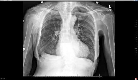
The plain film radiograph of the chest demonstrated a fluid level (yellow arrow) in the distal esophagus with dilation of the esophagus proximal to that point (blue line). Neither of these findings were present on the previous visit.
Abdominal/GastroenterologyVisual EM
Creative Commons images

Case Report: Talar Neck Fracture

Wilson Frasca, MD* and Nhan Do, MD*

DOI: https://doi.org/10.21980/J8FP75 Issue 5:3[mrp_rating_result]
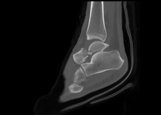
ABSTRACT: This report demonstrates a case of a severe talar neck fracture. Although rare, talar neck fractures have a high potential for morbidity. Typically caused by a high energy injury, this patient’s mechanism of injury was relatively minor, and presentation was not immediately concerning for such a severe fracture. Initial x-rays provided a gross demonstration of the fracture, but a
OrthopedicsVisual EM
Creative Commons images

A Case Report on Miliary Tuberculosis in Acute Immune Reconstitution Inflammatory Syndrome

Erica Concors, MD*, Hamid Ehsani-Nia, DO* and Michael Mirza, MD*

DOI: https://doi.org/10.21980/J81H02 Issue 5:3[mrp_rating_result]
A portable single-view radiograph of the chest was obtained upon the patient’s arrival to the ED resuscitation bay that showed diffuse reticulonodular airspace opacities (red arrows) seen throughout the bilateral lungs, concerning for disseminated pulmonary tuberculosis. Subsequently, a computed tomography (CT) angiography of the chest was obtained which again demonstrates this diffuse reticulonodular airspace opacity pattern (red arrows).
Infectious DiseaseRespiratoryVisual EM
Creative Commons images

Case Report of Untreated Pediatric Femoral Neck Fracture with Osteopenia

Sha Yan, DO*

DOI: https://doi.org/10.21980/J8S92K Issue 5:2[mrp_rating_result]
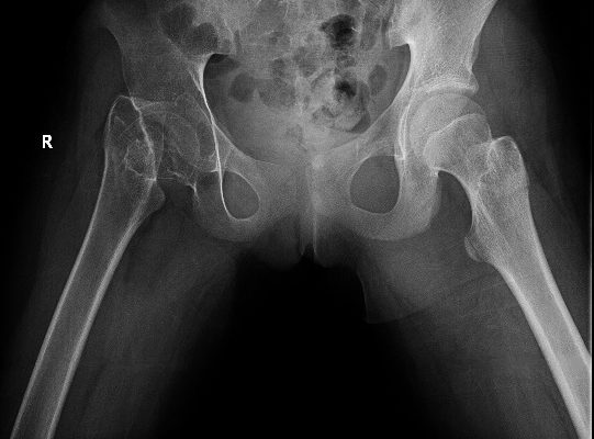
On her right hip radiograph, the patient was found to have a right femoral neck fracture with superior displacement of the intertrochanteric portion of the right femur. Moreover, the radiograph demonstrated diffuse osteopenia of the right hip and femur from chronic disuse as characterized by the increased radiolucency of the cortical bones compared to the left side.
OrthopedicsPediatrics
Creative Commons images

Loose PEG Tube Leading to Peristomal Leakage and Peritonitis

Connie Au, BS and Toby Myatt, MD

DOI: https://doi.org/10.21980/J8HS7T Issue 5:2[mrp_rating_result]
Frontal chest X-ray showed a large radiolucent area (pink highlighted area) underneath the diaphragm (yellow line) and on top of the liver (blue highlighted area) and spleen (green highlighted area) suggestive of pneumoperitoneum possibly caused by gastrointestinal perforation. This large radiolucent area can also be seen underneath the diaphragm in the lateral view chest X-ray. Computed tomography (CT) was not performed due to his physical exam findings and the significant positive findings on chest X-ray. Surgery was consulted and patient was taken emergently to the operating room.
Abdominal/GastroenterologyVisual EM
Creative Commons images

Rapid Airway Narrowing Associated with Hodgkin’s Lymphoma

Luke Hoffmann, BS* and Toby Myatt, MD*

DOI: https://doi.org/10.21980/J86D3Q Issue 5:2[mrp_rating_result]
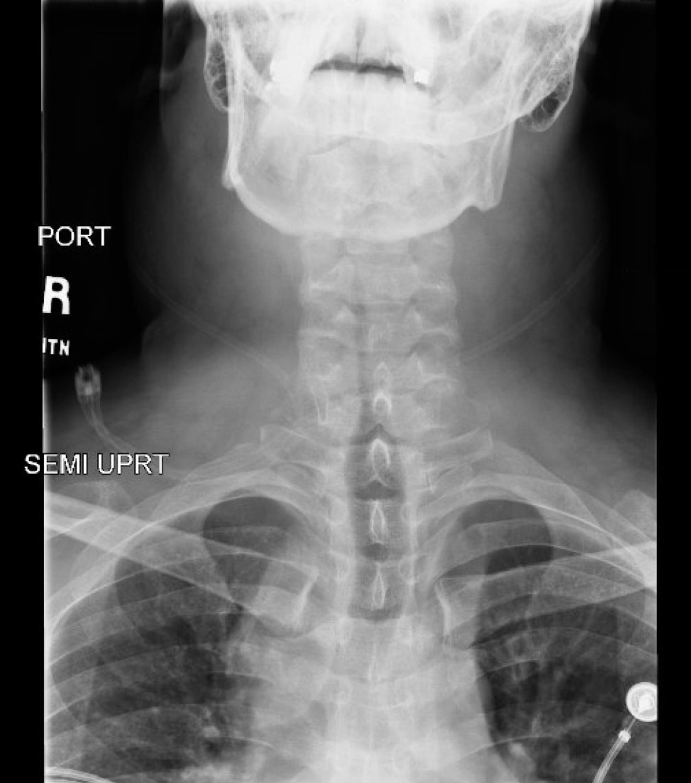
Neck X-ray showed nonspecific significant prevertebral soft tissue swelling at the level of the cervical spine, with associated apparent thickening of the epiglottis (yellow arrow), diffuse soft tissue swelling of the neck (red arrows) and tracheal airway narrowing (light blue arrow). The computed tomography imaging of the neck was significant for multiple conglomerating pathological lymph nodes with a significant mass effect (orange arrows) compressing the right internal jugular vein (green arrow).
Hematology/OncologyRespiratoryVisual EM
Creative Commons images

Ascending Thoracic Aortic Dissection: A Case Report of Rapid Detection Via Emergency Echocardiography with Suprasternal Notch Views

Brandon Backlund, MD*, Anastasia Kendrick-Adey, MD*, Rachel Harper, MD* and Martin Makela, MD*

DOI: https://doi.org/10.21980/J8WW6WIssue 5:2[mrp_rating_result]
Video of parasternal long-axis bedside transthoracic echocardiogram: The initial images showed grossly normal left ventricular function, and no pericardial effusion or evidence of cardiac tamponade. However, the proximal aorta beyond the aortic valve was poorly-visualized in this window.
Cardiology/VascularUltrasoundVisual EM
«‹4›»
Page 4 of 14

JETem is an online, open access, peer-reviewed, journal-repository for EM educators. We are PMC Indexed.

Most Viewed

  • Simple Auricular Hematoma (“Cauliflower Ear”) Task Trainer
  • The Three Breath Method: Teaching Coping Mechanisms for Processing Patient Death
  • Clinical Decision Making: Acute Ischemic Stroke with Large Vessel Occlusion
  • PRIMER: Premedical Introduction to Mentored Emergency Research: Connecting Undergraduate Assistants and Emergency Medicine Residents in a Community Hospital Setting
  • Dangerous Dysrhythmias in the ED: An Interactive EKG Workshop for Emergency Medicine Clerkship Learners

Visit Our Collaborators


Creative Commons Licence
This work is licensed under a Creative Commons Attribution 4.0 International License.

About

Education

Learners should benefit from active learning. JETem accepts submissions of team-based learning, small group learning, simulation, podcasts, lectures, innovations, curricula, question sets, and visualEM.

Scholarship

We believe educators should advance through the scholarship of their educational work. JETem gives educators the opportunity to publish scholarly academic work so that it may be widely distributed, thereby increasing the significance of their results.

Links

  • Home
  • Aim and Scope
  • Current Issue
  • For Reviewers
  • Instructions for Authors
  • Contact Us

Newsletter

Sign up to receive updates from JETem regarding newly published issues and findings.

Copyright Creative Commons Attribution 4.0 International