Photograph
Brown Recluse Spider Bite
DOI: https://doi.org/10.21980/J8TK99Examination of the skin revealed erythema and induration on the right lateral leg 12 cm in diameter with a 6 cm area of central necrosis and surrounding petechiae without fluctuance or crepitus. The patient was neurovascularly intact distal to the lesion. Laboratory values were within normal limits, except for an elevated C-reactive protein (5.31 mg/dL, normal range <0.70 mg/dL). The patient was diagnosed with ulceration secondary to envenomation from a brown recluse spider.
Levamisole Induced, Cocaine Associated Vasculitis
DOI: https://doi.org/10.21980/J8K35SAn asymmetric pattern of palpable purpura with bullae was noted on bilateral lower extremities with smaller patches on bilateral upper extremities. There was no tenderness or crepitus.
Pectoralis Muscle Tendon Rupture
DOI: https://doi.org/10.21980/J81D01There is a noticeable difference in appearance and location of maximal prominence of the right pectoral muscle with arms outstretched (image 1). This is accentuated by having the patient perform an isometric arm press. (image 2).There is absence of the anterior axillary fold with adduction against resistance. The stump of the pectoralis muscle was palpated along his armpit. He otherwise has full range of motion in the shoulder with minimal pain.
Abdominal Pain with Black Tongue
DOI: https://doi.org/10.21980/J8XS7JPatient’s tongue had a black discoloration, without elongated filiform papillae. We could not appreciate lymphadenopathy. His abdomen was tender to palpation.
Suspicious Skin Lesion in an 11-Year-Old Male
DOI: https://doi.org/10.21980/J8JK9TThe patient had a 5 cm ulcerative lesion with raised borders and a yellow, “fatty” center. There was no active drainage, site tenderness, or lymphadenopathy.
Warm & Blue: A Case of Methemoglobinemia
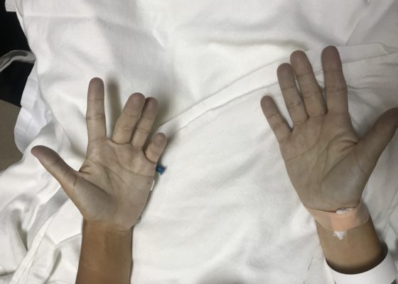
DOI: https://doi.org/10.21980/J8591MThe patient hadperioral cyanosis, blue coloration around her mouth, but the rest of the skin on her face appeared normal. She also had acrocyanosis to bilateral hands that can be seen in the image. The patient has a tan complexion up to the level of her wrists, but the palms of her hands are pale and cyanotic.
Open Globe with Intraocular Foreign Body
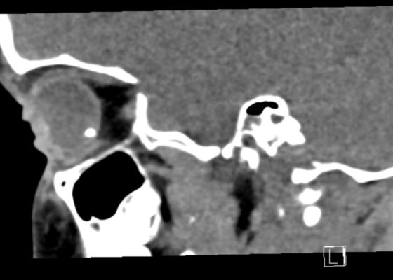
DOI: https://doi.org/10.21980/J8S348On physical exam, his extraocular movements were intact. The right anterior chamber appeared cloudy, particularly nasal to the pupil. The conjunctiva of the right eye was injected. The right pupil was 3 mm and sluggishly reactive and appeared slightly irregular (see yellow arrow). Of note, the right eye also had a 1 mm hypopyon, indicating inflammation of the anterior chamber, which was visible on slit lamp examination (not pictured). There was no fluorescein uptake or Seidel sign. His visual acuity was 20/60 OD (right eye) and 20/20 OS (left eye).
Hypopyon
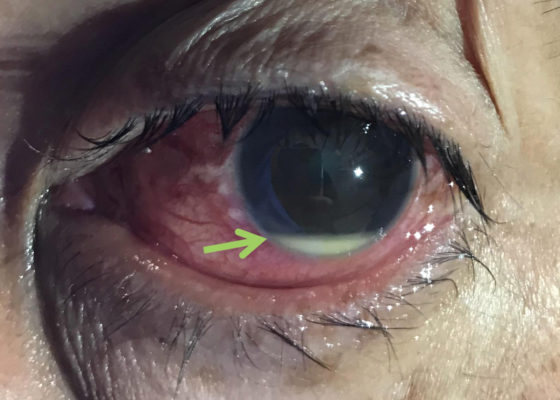
DOI: https://doi.org/10.21980/J8N92BPhysical examination of the left eye revealed a hypopyon (green arrow) – which is a layered white to yellow sediment in front of the inferior aspect of the iris associated with scleral injection and chemosis. Extraocular movements were intact bilaterally and pain did not worsen with extraocular movement. The pupil was poorly reactive to direct light and only hand movement could be perceived. The intraocular pressure was 14 mmHg. Slit lamp exam demonstrated a dense cataract. Bedside ocular ultrasound demonstrated vitreous opacities concerning for possible intraocular foreign bodies.


