Traumatic Hyphema
History of present illness:
A 40-year-old male presented to the emergency department with report of being shot in the right eye by a foam dart gun just prior to arrival. He was unable to see out of the affected eye. He initially had some pain in the eye, but it had subsided prior to arrival.
Significant findings:
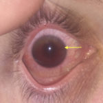
Upon initial evaluation, the patient had an obvious hyphema in the right eye with associated conjunctival injection. Initially, the bleeding in the anterior chamber was cloudy just above the level of the pupil (yellow arrow), appearing to possibly be a grade II hyphema. There were no other signs of trauma to the eye under Wood’s lamp examination with fluorescein staining. The globe was intact. Intraocular pressure in the affected eye was 19 mmHg and 15 mmHg in the unaffected eye. Extraocular movements were full and intact. The pupil was 4 mm round and reactive to direct and consensual light. Visual acuity was greater than 20/200 in the affected eye compared to 20/25 in the unaffected eye. After an observation period of two hours, with the patient remaining upright, the hyphema had settled down to a rim in the lower anterior chamber (green arrow), a grade I hyphema.
Discussion:
A hyphema is a collection of blood in the anterior chamber of the eye. Most cases are the result of direct trauma and subsequent bleeding from the ciliary vessels. The incidence of traumatic hyphema is estimated to be 12 per 100,000 with males composing nearly eighty percent of cases.1 Rare atraumatic causes can be attributed to intraocular malignancy, bleeding disorders or antiplatelet and anticoagulant medications.2,3 A hyphema is graded based on the depth of blood in the anterior chamber. A grade I hyphema involves less than one-third of the anterior chamber, grade II is defined as one-third to one-half of the anterior chamber, grade III is greater than one-half of the anterior chamber, and grade IV is a total or “8 ball” hyphema. A microphyphema is only visible on slit lamp examination as floating red blood cells. Visual prognosis is related to the degree of hyphema, with a grade I hyphema having a 10 percent chance of vision worse than 20/50 compared to a grade III hyphema having a 50 to 75 percent chance of vision worse than 20/50.4,5
This case illustrates that on initial presentation a hyphema may appear to be graded higher and that a short observation period with the patient seated upright is required to provide an accurate diagnosis and prognosis. The second image also demonstrates the subtleness of a grade I hyphema. Recognition of a smaller hyphema is important because while the lower grade implies better prognosis, complications such as re-bleeding or uncontrolled intraocular pressure may still occur.5 The patient in this case had improvement of his vision throughout observation. He was discharged home with atropine ophthalmic drops and close ophthalmologic follow up.
Topics:
Traumatic hyphema, ophthalmology.
References:
- Kennedy RH, Brubaker RF. Traumatic hyphema in a defined population. Am J Ophthalmol. 1988;106(2):123. doi: 10.1016/j.ajo.2016.01.003
- Winegarner A, Hashida N, Koh S, Nishida K. Hemorrhagic hypopyon as presenting feature of intravascular lymphoma, a case report. BMC Ophthalmol. 2017;17(1):195. doi: 10.1186/s12886-017-0591-3
- Shieh W, Sridhar J, Hong BK, et al. Ophthalmic complications associated with direct oral anticoagulant medications. Semin Ophthalmol. 2017; 32(5):614-619. doi: 10.3109/08820538.2016.1139738
- Brandt MT and Haug RH. Traumatic hyphema: a comprehensive review. J Oral Maxillofac Surg. 2001;59(12):1462. doi: 10.1053/joms.2001.28284.
- Walton W, Von Hagen S, Grigorian R, Zarbin M. Management of traumatic hyphema. Surv Opthalmol. 2002;47(4):297-394.
- Guluma K, Lee J. Opthalmology. In: Walls RM, Hockberger RS, Gausche-Hill M, et al. eds Rosen’s Emergency Medicine: Concepts and Clinical Practice. 9th Philadelphia, PA: Elsevier; 2018:790-819.
- Ostermayer D, Westafer L, et al. Traumatic WikEM. https://wikem.org/wiki/Traumatic_hyphema. Published November 2017. Accessed June 9, 2018.
- Nickson C. Half an 8 ball. Life in the Fast Lane. https://lifeinthefastlane.com/ophthalmology-befuddler-030/. Published May 2016. Accessed June 9, 2018.


