• Registration
  • Login
JETem
  • Home
  • About
    • Aim and Scope
    • Our Team
    • Editorial Board
    • FAQ
  • Issues
    • Current Issue
    • Ahead of Print
    • Past Issues
  • Visual EM
    • Latest Visual EM
    • Search Visual EM
    • Thumbnail Library
  • For Authors
    • Instructions for Authors
    • Submit to JETem
    • Photo Consent
    • Policies
      • Peer Review Policy
      • Copyright Policy
      • Editorial Policy, Ethics and Responsibilities
      • Conflicts of Interest & Informed Consent
      • Open Access Policy
  • For Reviewers
    • Instructions for JETem Reviewers
    • Interested in Being a JETem Reviewer?
  • Topic
    • Abdominal / Gastroenterology
    • Administration
    • Board Review
    • Cardiology / Vascular
    • Clinical Informatics, Telehealth and Technology
    • Dermatology
    • EMS
    • Endocrine
    • ENT
    • Faculty Development
    • Genitourinary
    • Geriatrics
    • Hematology / Oncology
    • Infectious Disease
    • Miscellaneous
    • Neurology
    • Ob / Gyn
    • Ophthalmology
    • Orthopedics
    • Pediatrics
    • Pharmacology
    • Procedures
    • Psychiatry
    • Renal / Electrolytes
    • Respiratory
    • Social Determinants of Health
    • Toxicology
    • Trauma
    • Ultrasound
    • Urology
    • Wellness
    • Wilderness
  • Modality
    • Curricula
    • Innovations
    • Lectures
    • Oral Boards
      • Structured Interview
      • Communication Case
    • Podcasts
    • Simulation
    • Small Group Learning
    • Team Based Learning
    • Visual EM
  • Contact Us

Issue 7:3

Zombie Cruise Ship Virtual Escape Room for POCUS Pulmonary: Scan Your Way Out

Heesun Choi, DO*^, Alisa Wray, MD, MAEd* and Jonathan Smart, MD *

DOI: https://doi.org/10.21980/J8RM0MIssue 7:3 No ratings yet.
By the end of performing the Zombie Cruise Ship Virtual Escape Room, learners will be able to: 1) recognize sonographic signs of A-line, B-line, Barcode sign, Bat sign, Seashore Sign, Plankton sign, Jellyfish Sign, Lung point, lung lockets, and Lung pulse; 2) differentiate sonographic findings of pneumothorax, hemothorax, pneumonia, COVID 19 pneumonia, pulmonary edema, and pleural effusion from normal lung findings; 3) distinguish pneumonia from atelectasis by recognizing dynamic air bronchogram; and 4) recognize indications for performing POCUS pulmonary such as dyspnea, blunt trauma, fall, cough and/or heart failure. 
UltrasoundRespiratorySmall Group Learning

High-Efficiency Ultrasound-Guided Regional Nerve Block Workshop for Emergency Medicine Residents

Brandon Yonel, BS*, Eunice Kwak, BS* and Mohamad Moussa, MD, RDMS*

DOI: https://doi.org/10.21980/J84P8R Issue 7:3 No ratings yet.
The objective of this workshop is to provide emergency medicine residents the confidence and skill sets needed to effectively perform five commonly used UGRNBs for conditions encountered in the emergency department. Through this one-day, accelerated workshop, residents will be given an opportunity to sharpen their UGRNB technique prior to applying them in the clinical environment. By the end of this workshop, learners will be able to: 1) recognize the clinical situations in which UGRNBs can be utilized and understand the associated risks, 2) list the commonly used local anesthetic medications and their proper dosing in respect to regional nerve blocks, 3) demonstrate proper ultrasound probe positioning and identify relevant anatomical landmarks for each nerve block on both standardized patients and cadavers, 4) describe the common steps involved to perform each nerve block, 5) perform the five UGRNB techniques outlined in this workshop.
ProceduresSmall Group LearningUltrasound

Cyanide Poisoning

Ghadeer Doman, MD*, Jihad Aoun, MS^, Joshua Truscinski, MS^, Mariah Truscinski, MD^ and Shaza Aouthmany, MD^

DOI: https://doi.org/10.21980/J80W76 Issue 7:3 No ratings yet.
After the completion of this simulation, participants will have learned how to: 1) identify clues of smoke inhalation based on a physical examination; 2) identify smoke inhalation-induced airway compromise and perform definitive management; 3) create a differential diagnosis for victims of fire cyanide poisoning, carbon monoxide, and carbon dioxide; 4) appropriately treat cyanide poisoning; 5) demonstrate the importance of preemptively treating for cyanide poisoning; 6) perform an initial physical examination and identify physical marks suggesting the patient is a fire and smoke inhalation victim; and 7) familiarize themselves with the Cyanokit and treatment with hydroxocobalamin.
ToxicologySimulation

Aortic Dissection Presenting as a STEMI

Jennifer Yee, DO* and Andrew P Kendle, MD*

DOI: https://doi.org/10.21980/J8W647 Issue 7:3 No ratings yet.
At the conclusion of the simulation session or during the debriefing session, learners will be able to: 1) Verbalize the anatomical differences and management of Stanford type A and type B aortic dissections, 2) Describe physical exam findings that may be found with ascending aortic dissections, 3) Describe the various clinical manifestations of the propagation of aortic dissections, 4) Discuss the management of aortic dissection, including treatment and disposition.
Cardiology/VascularSimulation

Morphine Equianalgesic Dose Chart in the Emergency Department

Savannah Tan, MD*, Ellen Lee, PharmD*, Stephen Lee, PharmD*, Sangeeta S Sakaria, MD, MPH, MST* and Jennifer S Roh, MD*

DOI: https://doi.org/10.21980/J8RD29 Issue 7:3 No ratings yet.
By the end of this session, the learner will be able to: 1) define the term, “morphine milligram equivalents;” 2) describe the relative onset and duration of action of different pain medications often used in the emergency department; and 3) convert one opioid dose to another.
PharmacologyLectures

Acute Flaccid Myelitis

Dane Zappa, MD* and Linda L Herman, MD, FACEP^

DOI: https://doi.org/10.21980/J8MP9G Issue 7:3 No ratings yet.
At the end of this oral board session, examinees will: 1) demonstrate the ability to obtain a complete pediatric medical history; 2) demonstrate an appropriate exam on a pediatric patient including a neurological exam; 3) investigate the broad differential diagnoses for neuromuscular weakness in a pediatric patient; 4) order the appropriate evaluation studies including an MRI; 5) interpret the use of a negative inspiratory force in determining the need for intubation and level of care upon admission; and 6) demonstrate effective communication with parents and caregivers.
NeurologyOral Boards

Residents Are Coming: A Faculty Development Curriculum to Prepare a Community Site For New Learners

Keith Willner, MD*, Essie Reed-Schrader, MD* and Stephen Mohney, MD*

DOI: https://doi.org/10.21980/J87D2N Issue 7:3 No ratings yet.
Our goal is to prepare community-based EM attendings to be outstanding educators to future residents by augmenting their knowledge of current educational practice and adult learning theory, literature review, and biostatistics.
Faculty DevelopmentAdministrationCurricula
Creative Commons images

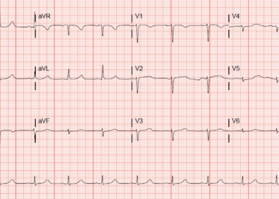
‘Cath’ It Before It’s Too Late: A Case Report of ECG Abnormalities Indicative of Acute Pathology Requiring Immediate Catheterization

Diane Wei, BS*, Paul Truong, DO*^ and Patrick Bruss, MD*^

DOI: https://doi.org/10.21980/J8HW7V Issue 7:3 No ratings yet.
A 12 lead ECG performed at the time of emergency department (ED) admission revealed regular sinus rhythm with noted T-wave inversion (blue arrows on Figure 1) in Lead aVL new when compared to an ECG performed a few months prior (see Figure 3). Two days later a second ECG was done when the patient developed acute chest pain while in the ICU (Figure 2) that showed persistent inversion in Lead aVL (blue arrows) as well as new J point deviation (JPD) in Leads II, aVF, V5 and V6; and new JPD in Leads V1 and V2 (green arrows) from her previous ECG while in the emergency department. These focal repolarization abnormalities did not qualify as an ST-elevation myocardial infarction by current guidelines.
Cardiology/VascularVisual EM
Creative Commons images

Evaluation of ACE-inhibitor Induced Laryngeal Edema Using Fiberoptic Scope: A Case Report

Joya Singh, DO* and Colin Danko, MD *

DOI: https://doi.org/10.21980/J83P9T Issue 7:3 No ratings yet.
Physical exam was initially significant for swelling isolated to the right sided cheek and upper lip. There was no edema to lower lip, uvular swelling, or swelling to the submandibular space. She was speaking full sentences and did not endorse any voice changes. Initial vital signs were as follows: BP 125/77, HR 74, RR 16, and oxygen saturation of 100% on room air. Approximately 40 minutes later, after 125 mg solumedrol intravenous (IV) and 50mg diphenhydramine by mouth, swelling had spread to the entire upper lip and the patient reported spreading to her jaw (Photo 1). Although no jaw or submandibular edema was appreciated on physical exam, a flexible fiberoptic laryngoscope was used to evaluate the patient’s airways given worsening symptoms. Viscous lidocaine was applied intranasally five minutes prior to the procedure. The patient was positioned in a seated position on the stretcher. A flexible fiberoptic laryngoscope was then inserted through the nares and advanced slowly. Laryngoscopy showed diffuse edema of the epiglottis, arytenoids, and ventricular folds (see photos 2-4). Vital signs and respiratory status remained stable both during and after the procedure.
ENTProceduresVisual EM
Creative Commons images

A Case Report of May-Thurner Syndrome Identified on Abdominal Ultrasound

Michelle Brown, DO*, Edmund Hsu, MD*, Christopher McCoy, MD* and Matthew Whited, MD*

DOI: https://doi.org/10.21980/J8C64K Issue 7:3 No ratings yet.
The patient initially received a venous doppler ultrasound that showed no evidence of a right or left femoropopliteal venous thrombus. Due to the high suspicion of a DVT given the symmetric swelling to the entire limb and acute onset of pain, a CTV was ordered. The transverse view of the CTV showed chronic thrombotic occlusion of the proximal left common iliac vein associated with compression from the right common iliac artery (figure 1, transverse image of CTA), as well as thrombotic occlusion of the left internal iliac vein tributary and corresponding left ascending lumbar vein. Given the previously mentioned clinical context, these features suggested the diagnosis of May-Thurner syndrome.
Cardiology/VascularVisual EM
1›
Page 1 of 2

JETem is an online, open access, peer-reviewed, journal-repository for EM educators. We are PMC Indexed.

Most Viewed

  • The Silent Saboteur: Teaching the Clinical Implications of Occult Hypoxemia & Social Determinants of Health via a Pulmonary Embolism Case
  • Diabetic Ketoacidosis and Necrotizing Soft Tissue Infection
  • My Broken Heart
  • Stabilization of Cardiogenic Shock for Critical Care Transport, a Simulation
  • Innovative Ultrasound-Guided Erector Spinae Plane Nerve Block Model for Training Emergency Medicine Physicians

Visit Our Collaborators

Creative Commons Licence
This work is licensed under a Creative Commons Attribution 4.0 International License.

About

Education

Learners should benefit from active learning. JETem accepts submissions of team-based learning, small group learning, simulation, podcasts, lectures, innovations, curricula, question sets, and visualEM.

Scholarship

We believe educators should advance through the scholarship of their educational work. JETem gives educators the opportunity to publish scholarly academic work so that it may be widely distributed, thereby increasing the significance of their results.

Links

  • Home
  • Aim and Scope
  • Current Issue
  • For Reviewers
  • Instructions for Authors
  • Contact Us

Newsletter

Sign up to receive updates from JETem regarding newly published issues and findings.

Copyright Creative Commons Attribution 4.0 International