• Registration
  • Login
JETem
  • Home
  • About
    • Aim and Scope
    • Our Team
    • Editorial Board
    • FAQ
  • Issues
    • Current Issue
    • Ahead of Print
    • Past Issues
  • Visual EM
    • Latest Visual EM
    • Search Visual EM
    • Thumbnail Library
  • For Authors
    • Instructions for Authors
    • Submit to JETem
    • Photo Consent
    • Policies
      • Peer Review Policy
      • Copyright Policy
      • Editorial Policy, Ethics and Responsibilities
      • Conflicts of Interest & Informed Consent
      • Open Access Policy
  • For Reviewers
    • Instructions for JETem Reviewers
    • Interested in Being a JETem Reviewer?
  • Topic
    • Abdominal / Gastroenterology
    • Administration
    • Board Review
    • Cardiology / Vascular
    • Clinical Informatics, Telehealth and Technology
    • Dermatology
    • EMS
    • Endocrine
    • ENT
    • Faculty Development
    • Genitourinary
    • Geriatrics
    • Hematology / Oncology
    • Infectious Disease
    • Miscellaneous
    • Neurology
    • Ob / Gyn
    • Ophthalmology
    • Orthopedics
    • Pediatrics
    • Procedures
    • Psychiatry
    • Renal / Electrolytes
    • Respiratory
    • Toxicology
    • Trauma
    • Ultrasound
    • Wellness
    • Wilderness
  • Modality
    • Curricula
    • Innovations
    • Lectures
    • Oral Boards
    • Podcasts
    • Simulation
    • Small Group Learning
    • Team Based Learning
    • Visual EM
  • Contact Us

A Case Report of May-Thurner Syndrome Identified on Abdominal Ultrasound

Michelle Brown, DO*, Edmund Hsu, MD*, Christopher McCoy, MD* and Matthew Whited, MD*

*University of California, Irvine, Department of Emergency Medicine, Orange, CA

Correspondence should be addressed to Michelle Brown, DO at mdbrown1@hs.uci.edu

DOI: https://doi.org/10.21980/J8C64K Issue 7:3
Cardiology/VascularVisual EM
No ratings yet.

ABSTRACT:

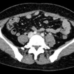
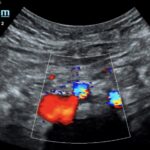
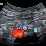
May-Thurner syndrome (MTS) is most commonly caused by the compression of the left iliac vein by the right iliac artery against the lumbar spine, which leads to the development of a partial or occlusive deep venous thrombosis (DVT).1 Diagnosis begins with a duplex ultrasound of the lower extremities to evaluate for a femoropopliteal thrombus, and in high-risk patients where a more proximal DVT is suspected and the DVT ultrasound is negative, a computed tomography venogram (CTV) or magnetic resonance venogram (MRV) is performed.1,3 In this case report, a patient presented to the emergency department (ED) with two days of left lower extremity pain and swelling. Initial lower extremity DVT ultrasound was negative, so a CTV was ordered and revealed a thrombus in the left common iliac vein with overlying compression by the right common iliac artery, suggesting the diagnosis of May-Thurner syndrome (Figure 1). Afterwards, a point-of-care ultrasound (POCUS) was performed at bedside to evaluate the caval and iliac arteries and the findings were consistent with the CTV (Figure 2, 3, 4). If the POCUS was performed prior to the CTV, the patient may have been spared the radiation exposure from CT, as well as the risks of intravenous (IV) contrast required for a venogram. Therefore, in high risk patients in whom a negative DVT ultrasound will prompt advanced imaging with CTV or MRV, I propose the addition of a lower abdominal ultrasound using a curvilinear probe to assess the caval and iliac arteries prior to obtaining a CTV or MRV.

Topics:

May-Thurner Syndrome, leg swelling, POCUS, ultrasound, deep venous thrombosis.

Icon

May-Thurner Syndrome - Manuscript

1 file(s) 3.96 MB
Download
Icon

May-Thurner Syndrome - Images

1 file(s) 2.17 MB
Download
CTIssue 7:3Ultrasound

Reviews:

No ratings yet.

Please rate this


Vitreous Hemorrhage Case Report

30 Jun, 22

Evaluation of ACE-inhibitor Induced Laryngeal...

30 Jun, 22
JETem is an online, open access, peer-reviewed journal-repository for EM educators

Most Viewed

  • A Model Curriculum for an Emergency Medicine Residency Rotation in Clinical Informatics
  • Use of An Ophthalmology Tutorial to Improve Resident Comfort with the Emergency Eye Exam
  • A Novel Module Based Method of Teaching Electrocardiogram Interpretation for Emergency Medicine Residents
  • Respiratory Distress in the Pediatric ED: A Case-based Self-directed Learning Module
  • Methemoglobinemia

Visit Our Collaborators