Issue 4:2
Acute Ischemic Stroke
DOI: https://doi.org/10.21980/J8R04XBy the end of this simulation session, learners will be able to: 1) recognize a CVA using the National Institutes of Health Stroke Scale (NIHSS), 2) understand and properly utilize the NIHSS, 3) list appropriate imaging and laboratory orders for a CVA work-up, 4) determine appropriate subspecialty consultation, 5) discuss common stroke syndromes and associated cerebral locations, 6) review indications and contraindications for tissue plasminogen activator (tPA), 7) review hospital specific stroke protocol.
Ethylene Glycol Ingestion
DOI: https://doi.org/10.21980/J8M620By the conclusion of the simulation session, learners will be able to: 1) obtain a thorough toxicologic history, including intent, timing, volume/amount, and assessment for co-ingestions, 2) distinguish the variable clinical signs and symptoms associated with toxic alcohol ingestions, 3) identify metabolic derangements associated with toxic alcohol ingestions, 4) discuss the management of toxic alcohol ingestion, 5) appropriately disposition the patient for admission to intensive care unit (ICU).
Approach to Geriatric Emergency Medicine: A Flipped Classroom Group Learning Exercise for Undergraduate Medical Trainees
DOI: https://doi.org/10.21980/J8GH03At the end of the module, learners should be able to: 1) recognize that many benign-seeming presentations, including restricting fatigue and cognitive decline, can have serious and life-threatening causes, 2) describe the importance of screening for delirium in older ED patients, 3) identify situations in which vital signs can be misleading in older adults and know strategies to further investigate such data, and 4) recognize that older adults can rapidly develop delirium in the ED and be able to apply strategies to reduce risk of delirium.
Emergency Medicine Curriculum Utilizing the Flipped Classroom Method: Environmental Emergencies
DOI: https://doi.org/10.21980/J8TP8ZThrough a flipped classroom design, we aim to teach the presentation and management of environmental emergencies, specifically cold related illness, heat related illness, undersea medicine, high altitude medicine, submersion, electrocution, radiation injury, and envenomation. This unique, innovative curriculum utilizes resources chosen by education faculty and resident learners, study questions, real-life experiences, and small group discussions in place of traditional lectures. In doing so, a goal of the curriculum is to encourage self-directed learning, improve understanding and knowledge retention, and improve the educational experience of our residents.
Google Forms – A Novel Solution to Blended Learning
DOI: https://doi.org/10.21980/J8BP77By the end of the session, the learner should be able to create a didactic session utilizing Google Forms (or similar web-based application). Specific learning objectives for the didactic session will vary based on application. Our institution has used Google Forms to create case-based small group discussion sessions, “create your own adventure” individual learning cases, asynchronous learning opportunities, and interactive intra-lecture surveys.
The Gravid Watermelon: An Inexpensive Perimortem Caesarean Section Model
DOI: https://doi.org/10.21980/J8705NThe gravid watermelon is a cost-effective model that uses common materials from the supermarket and emergency department (ED), using a carved-out watermelon as a base, representing the peritoneal cavity. Inexpensive respiratory tubing is used to represent intestine; watered down gelatin and a small doll in a deflated rubber/plastic ball is used to represent a gravid uterus. The bladder is represented by an unused, water-filled exam glove, and watermelon pulp represents blood clots and mesentery. The gravid watermelon is covered with an elastic bandage to represent tough muscle and fascia, and topped with a shower curtain for skin.
Low-Cost Portable Suction-Assisted Laryngoscopy Airway Decontamination (SALAD) Simulator for Dynamic Emesis
DOI: https://doi.org/10.21980/J8362BThe economic and dynamic SALAD innovation recreates an actively vomiting patient and replicates visual obstruction from fluid contents during airway management.By the end of the session, learners are expected to: 1) discuss the risks, benefits, indications and contraindications associated with intubation of a vomiting or hemorrhaging patient. 2) Work with colleagues to effectively stabilize a patient who is actively vomiting or bleeding during airway management. 3) Competently perform intubation in the acute setting of visual obstruction from active emesis, hemorrhage, or massive regurgitation. 4) Increase speed and dexterity of intubation by applying the SALAD method when fluid obstructs visualization of the larynx.
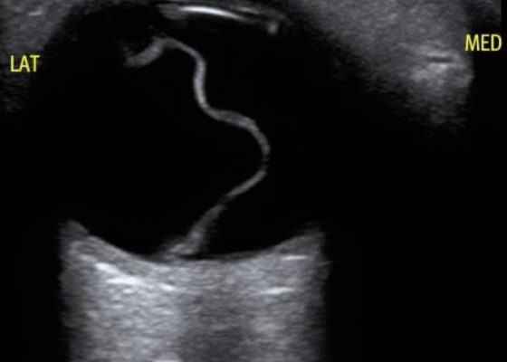
Macula-Off Retinal Detachment Identified on Bedside Ultrasound
DOI: https://doi.org/10.21980/J8WP8KPoint-of-care ultrasound was performed, demonstratinga free-floating, serpiginous, hyperechoic membrane (R) tethered at the optic nerve (ON) and ora serrata (OS), but detached at the macula (M) lateral to the optic nerve. This is diagnostic for macula-off retinal detachment. It can be differentiated from macula-on retinal detachment, in which the hyperechoic retina would appear attached posteriorly at the location of the macula just lateral to the optic nerve. Ophthalmology was consulted, agreed with the diagnosis of macula-off retinal detachment, and took the patient to the OR for laser photocoagulation.

