• Registration
  • Login
JETem
  • Home
  • About
    • Aim and Scope
    • Our Team
    • Editorial Board
    • FAQ
  • Issues
    • Current Issue
    • Ahead of Print
    • Past Issues
  • Visual EM
    • Latest Visual EM
    • Search Visual EM
    • Thumbnail Library
  • For Authors
    • Instructions for Authors
    • Submit to JETem
    • Photo Consent
    • Policies
      • Peer Review Policy
      • Copyright Policy
      • Editorial Policy, Ethics and Responsibilities
      • Conflicts of Interest & Informed Consent
      • Open Access Policy
  • For Reviewers
    • Instructions for JETem Reviewers
    • Interested in Being a JETem Reviewer?
  • Topic
    • Abdominal / Gastroenterology
    • Administration
    • Board Review
    • Cardiology / Vascular
    • Clinical Informatics, Telehealth and Technology
    • Dermatology
    • EMS
    • Endocrine
    • ENT
    • Faculty Development
    • Genitourinary
    • Geriatrics
    • Hematology / Oncology
    • Infectious Disease
    • Miscellaneous
    • Neurology
    • Ob / Gyn
    • Ophthalmology
    • Orthopedics
    • Pediatrics
    • Pharmacology
    • Procedures
    • Psychiatry
    • Renal / Electrolytes
    • Respiratory
    • Social Determinants of Health
    • Toxicology
    • Trauma
    • Ultrasound
    • Urology
    • Wellness
    • Wilderness
  • Modality
    • Curricula
    • Innovations
    • Lectures
    • Oral Boards
      • Structured Interview
      • Communication Case
    • Podcasts
    • Simulation
    • Small Group Learning
    • Team Based Learning
    • Visual EM
  • Contact Us

Issue 3:2

Creative Commons images

Necrotizing Soft Tissue Infection

Sahil Aggarwal, BS* and Jonathan Peña, MD*

DOI: https://doi.org/10.21980/J8X92TIssue 3:2 No ratings yet.
Computed tomography (CT) of the abdominal and pelvis with intravenous (IV) contrast revealed inflammatory changes, including gas and fluid collections within the ventral abdominal wall extending to the vulva, consistent with a necrotizing soft tissue infection.
Infectious DiseaseVisual EM
Creative Commons images

Spontaneous Pneumomediastinum: Hamman Syndrome

Tushank Chadha, BS* and Ethan Kunstadt, MD*

DOI: https://doi.org/10.21980/J8NS72 Issue 3:2 No ratings yet.
The initial CT scans showed extraluminal gas surrounding the distal esophagus as it traversed the posterior mediastinum, concerning for possible distal esophageal perforation that prompted surgery and GI consultations. There was no evidence of a drainable collection or significant fat stranding. The image also showed an intraluminal stent traversing the gastric antrum and gastric pylorus with no indication of obstruction. Circumferential mural thickening of the gastric antrum and body were consistent with the patient’s history of gastric adenocarcinoma. The shotty perigastric lymph nodes with associated fat stranding, along the greater curvature of the distal gastric body suggested local regional nodal metastases and possible peritoneal carcinomatosis. The thoracic CT scans showed extensive pneumomediastinum that tracked into the soft tissues of the neck, which given the history of vomiting also raised concern for esophageal perforation. There was still no evidence of mediastinal abscess or fat stranding. Additionally, a left subclavian vein port catheter, which terminates with tip at the cavoatrial junction of the superior vena cava can also be seen on the image.
RespiratoryVisual EM
Creative Commons images

Intussusception

Julian Jakubowski, DO*

DOI: https://doi.org/10.21980/J8SH0WIssue 3:2 No ratings yet.
A segment of bowel within the right abdomen that measured approximately 1.6 x 1.5 cm transaxially. It demonstrated a hypoechoic edematous outer loop of bowel (blue arrow) and hyperechoic compressed loop of bowel telescoping within (red star), this is known as the "target sign."
Abdominal/GastroenterologyVisual EM
Creative Commons images

Lateral Epicondyle Fracture

Valerie Lew, BS*, John Costumbrado, MD, MPH* and Curtis Knight, MD*

DOI: https://doi.org/10.21980/J8J05F Issue 3:2 No ratings yet.
Radiographs of the right elbow revealed an acute fracture through the lateral epicondyle with dislocation of the radial head inferiorly. Radiographs of the left elbow revealed a slightly angulated fracture through the lateral epicondyle.
OrthopedicsVisual EM
Creative Commons images

Propafenone Overdose-induced Arrhythmia and Subsequent Correction After Administration of Sodium Bicarbonate

Patrick Bruss, MD* and Peter Fuller, MD^

DOI: https://doi.org/10.21980/J8D925Issue 3:2 No ratings yet.
The first ECG in this case showed sinus tachycardia with a widened QRS (black arrow), a rightward axis, prolonged corrected QT interval (QTc), and terminal R wave in AVR (white arrow). There are several potential causes for these ECG findings, but put together with the patient’s history, we were suspicious of sodium channel blockers being the most likely cause. The second ECG, after sodium bicarbonate was administered, demonstrated a normal QRS (black arrow) and no rightward axis deviation, reduction of the QTC and resolution of the terminal R wave (white arrow).  We later learned that the patient’s cardiologist recently increased her propafenone dose.
ToxicologyVisual EM
Creative Commons images

Herpes Zoster Ophthalmicus

Shayna Adams, BS* and Darren Knight, MD^

DOI: https://doi.org/10.21980/J88H07 Issue 3:2 No ratings yet.
Physical exam was significant for an erythematous maculopapular rash in the right ophthalmic nerve (V1) region, a crusted vesicle at the right upper eyelid, and surrounding eyelid edema. Visual acuity remained at baseline and intraocular pressure was within normal limits. External slit lamp exam with fluorescein staining was remarkable for pseudodendrites in the inferior-temporal cornea approximately 1.5 mm from the limbus. Ophthalmology was consulted and completed an in-depth evaluation of the corneal lesions. They found no evidence of anterior chamber reaction and performed a complete dilated fundus examination which demonstrated no retinal involvement. The patient was then discharged on acyclovir and erythromycin ointment with close follow-up.
OphthalmologyVisual EM
Creative Commons images

Evaluation of Snake Bites with Bedside Ultrasonography

Josef E Jolissaint*, Alexis Salerno, MD^ and Daniel Sheets, MD^

DOI: https://doi.org/10.21980/J84S7D Issue 3:2 No ratings yet.
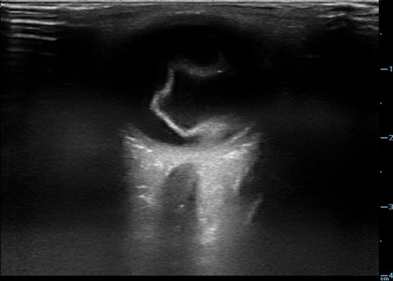
History of present illness: While watering his lawn, a 36-year-old man felt two sharp bites to his bilateral ankles. He reports that he then saw a light brown, 2-foot snake slither away from him. He came to the emergency department because of pain and swelling in his ankles and inability to bear weight. Physical examination revealed bilateral ankle swelling and
WildernessUltrasoundVisual EM
Creative Commons images

Procedural Sedation for the removal of a rectal foreign body

John Costumbrado, MD, MPH*, Valerie Lew, BS* and David Wagner, MD*

DOI: https://doi.org/10.21980/J81332Issue 3:2 No ratings yet.
Axial and coronal views on CT showed evidence of a large, tube-shaped foreign body in the rectum (see arrows) without evidence of acute gastrointestinal tract disease.
Abdominal/GastroenterologyProceduresVisual EM
Creative Commons images

Glass Foreign Body Hand Radiograph

Hamid Ehsani-Nia, DO* and Joshua Bucher, MD*

DOI: https://doi.org/10.21980/J8W92HIssue 3:2 No ratings yet.
History of present illness: A 27-year-old female sustained an injury to her left hand after she tripped and fell on a vase. She presented to the emergency department (ED) complaining of pain over the laceration. Upon examination, patient presented with multiple small abrasions of the medial aspect of the left 5thdigit that are minimally tender. Additionally, she has one 0.5cm
TraumaUltrasoundVisual EM
Creative Commons images

A Case of Otomastoiditis

Chad Correa, BS* and Claire Thomas, MD^

DOI: https://doi.org/10.21980/J8RK89Issue 3:2 No ratings yet.
The patient underwent computed tomography (CT) of the head which revealed opacification of the left middle ear (red arrow) and mastoid air cells (red circles). Additionally, there was thickening of the soft tissues of the external auditory canal (blue arrowhead), likely reflecting concurrent otitis externa. Based on the imaging, he was admitted for findings consistent with acute otomastoiditis.
ENTInfectious DiseaseVisual EM
‹2›»
Page 2 of 4

JETem is an online, open access, peer-reviewed, journal-repository for EM educators. We are PMC Indexed.

Most Viewed

  • The Silent Saboteur: Teaching the Clinical Implications of Occult Hypoxemia & Social Determinants of Health via a Pulmonary Embolism Case
  • Diabetic Ketoacidosis and Necrotizing Soft Tissue Infection
  • My Broken Heart
  • Stabilization of Cardiogenic Shock for Critical Care Transport, a Simulation
  • Innovative Ultrasound-Guided Erector Spinae Plane Nerve Block Model for Training Emergency Medicine Physicians

Visit Our Collaborators

Creative Commons Licence
This work is licensed under a Creative Commons Attribution 4.0 International License.

About

Education

Learners should benefit from active learning. JETem accepts submissions of team-based learning, small group learning, simulation, podcasts, lectures, innovations, curricula, question sets, and visualEM.

Scholarship

We believe educators should advance through the scholarship of their educational work. JETem gives educators the opportunity to publish scholarly academic work so that it may be widely distributed, thereby increasing the significance of their results.

Links

  • Home
  • Aim and Scope
  • Current Issue
  • For Reviewers
  • Instructions for Authors
  • Contact Us

Newsletter

Sign up to receive updates from JETem regarding newly published issues and findings.

Copyright Creative Commons Attribution 4.0 International