CT
Facial Fracture Induced Periorbital Emphysema
DOI: https://doi.org/10.21980/J8F05HPhysical exam showed marked left palpebral subcutaneous crepitus, as well as bulbar and palpebral conjunctival bulging. Visual acuity was normal with intact extraocular movements, and normal pupillary exam. Computed tomography (CT) imaging of the face was obtained and revealed multiple displaced fractures involving the left orbital floor and zygomatic arch associated with moderate periorbital and postseptal extraconal gas, resulting in orbital proptosis.
Fournier Gangrene
DOI: https://doi.org/10.21980/J89626The computed tomography (CT) of the abdomen and pelvis revealed significant subcutaneous gas tracking along the perineum and right gluteal region (orange outline) into the scrotum with associated scrotal edema (yellow arrow) and subcutaneous inflammatory fat stranding of 0.92 cm (red arrow) consistent with Fournier’s gangrene. There is early fluid loculation along the right medial gluteal cleft of 5.85 cm (green arrow) without a sizeable drainable abscess seen.
Foreign Body in Maxillary Sinus: A Rare Case of Chronic Rhinosinusitis
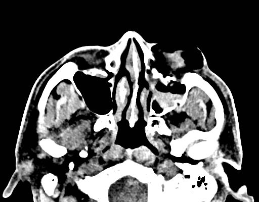
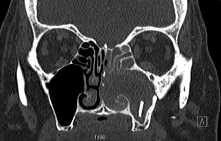
DOI: https://doi.org/10.21980/J85H09Computed tomography (CT) sinus with contrast demonstrated complete opacification of left paranasal sinuses and nasal cavity, and a linear radiopacity within the left maxillary sinus consistent with a foreign body. There were additional left facial subcutaneous radiopaque opacities.
Sialadenitis
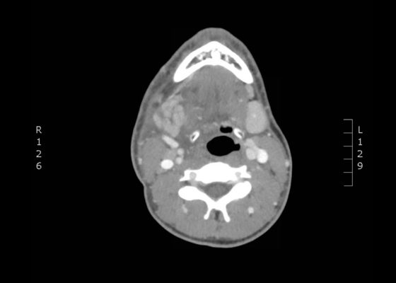
DOI: https://doi.org/10.21980/J8NH0NThe computed tomography (CT) scan demonstrates prominent enlargement and heterogeneous enhancement of the right submandibular gland (single large arrow) compatible with sialadenitis. There is no evidence of a sialolith or obstruction on the CT. There is associated edema (two small arrows) of the right submandibular space, parapharyngeal space and anterior right neck with partial effacement of the right vallecula and right pyriform sinus.
Subcutaneous Emphysema After Chest Trauma
DOI: https://doi.org/10.21980/J8864NPlain film anteroposterior (AP) radiography of the chest shows left-sided subcutaneous emphysema (red arrow) with overlapping muscle striations of the pectoralis major (green arrow). After chest tube placement (blue arrow), AP chest radiography shows persistent left-sided subcutaneous emphysema (red arrow). CT of the chest shows pneumomediastinum (blue arrow), left apical pneumothorax (pink arrow), and subcutaneous emphysema (red arrow) at the level of T2. At the level of T6, rib fractures can be visualized on the CT (yellow arrow). At the level of T8, left sided pneumothorax is also seen (pink arrow) as the absence of lung tissue on CT.
An Unusual Case of Hematemesis
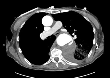
DOI: https://doi.org/10.21980/J84H00The patient’schest X-ray revealed a prominent mediastinum and opacification in the left middle and lower lung fields. The CT showed an aortic aneurysm extending from the thorax to the abdomen with rupture near T7 (blue arrow). It also showed periaortic hemorrhage with active extravasation (green arrow) likely secondary to a penetrating ulcer and bilateral pulmonary opacities concerning for hemothorax (pink arrow).
Extensive Aortic Dissection with Normal Vital Signs
DOI: https://doi.org/10.21980/J80S6SThe patient was found to have a Stanford type A dissection (see yellow arrow) with visible false lumen starting at aortic arch (see green circle). The dissection extended into the descending aorta (see blue circle) as shown by the false lumen (red highlighted area) visible on CT. The radiologist performed a reconstruction of the aorta, which showed that the left kidney was not being perfused, making the kidney not visible on the reconstruction.
Recurrent Sigmoid Volvulus in a Young Female
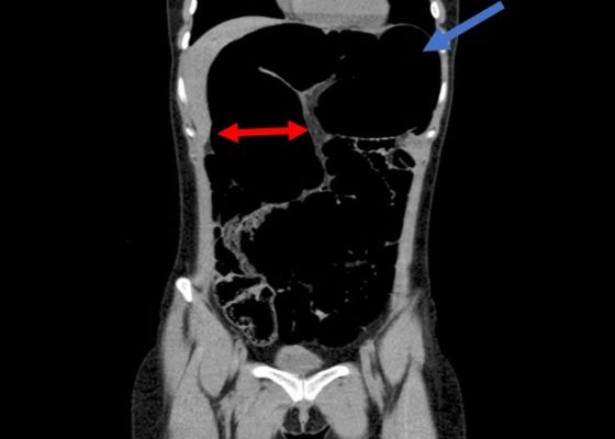
DOI: https://doi.org/10.21980/J8GW5SComputed tomography (CT) of the abdomen and pelvis was obtained revealing a colonic volvulus in the left mid to upper abdomen (blue arrow) involving the distal transverse colon and descending colon, with gaseous colonic distention to 8.5 cm (red arrow). The characteristic “whirl pattern” is also present (yellow arrow). These findings are suggestive of a high-grade colonic obstruction. It was without evidence of pneumoperitoneum, pneumatosis, or drainable collection. Of note, a 3.6 cm dermoid tumor is also observable in the left adnexa (green arrow).

