JETem
  • Home
  • About
    • Aim and Scope
    • Our Team
    • Editorial Board
    • FAQ
  • Issues
    • Current Issue
    • Ahead of Print
    • Past Issues
  • Visual EM
    • Latest Visual EM
    • Search Visual EM
    • Thumbnail Library
  • For Authors
    • Instructions for Authors
    • Submit to JETem
    • Photo Consent
    • Policies
      • Peer Review Policy
      • Copyright Policy
      • Editorial Policy, Ethics and Responsibilities
      • Conflicts of Interest & Informed Consent
      • Open Access Policy
      • Acceptance Fee
  • For Reviewers
    • Instructions for JETem Reviewers
    • Interested in Being a JETem Reviewer?
  • Topic
    • Abdominal/Gastroenterology
    • Administration
    • Board Review
    • Cardiology/Vascular
    • Certifying Exam Cases
      • Clinical Decision-Making
      • Prioritization
      • Communication
      • Procedure & Ultrasound
    • Clinical Informatics, Telehealth and Technology
    • Dermatology
    • EMS
    • Endocrine
    • ENT
    • Faculty Development
    • Genitourinary
    • Geriatrics
    • Hematology/Oncology
    • Infectious Disease
    • Miscellaneous (stats, etc)
    • Neurology
    • Ob/Gyn
    • Ophthalmology
    • Orthopedics
    • Pediatrics
    • Pharmacology
    • Procedures
    • Psychiatry
    • Renal/Electrolytes/Nutrition
    • Respiratory
    • Social Determinants of Health
    • Toxicology
    • Trauma
    • Ultrasound
    • Urology
    • Wellness
    • Wilderness
  • Modality
    • Certifying Exam Practice Cases
      • Clinical Care Cases
      • Communication & Procedural Cases
      • Old School Oral Board Cases
    • Curricula
    • Innovations
    • Lectures
    • Podcasts
    • Simulation
    • Small Group Learning
    • Team Based Learning (TBL)
    • Visual EM
  • Contact Us

CT

Creative Commons images

Subcutaneous Emphysema After Chest Trauma

Ronald Goubert, BS* and Alisa Wray, MD, MAEd*

DOI: https://doi.org/10.21980/J8864NIssue 3:4[mrp_rating_result]
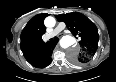
Plain film anteroposterior (AP) radiography of the chest shows left-sided subcutaneous emphysema (red arrow) with overlapping muscle striations of the pectoralis major (green arrow). After chest tube placement (blue arrow), AP chest radiography shows persistent left-sided subcutaneous emphysema (red arrow). CT of the chest shows pneumomediastinum (blue arrow), left apical pneumothorax (pink arrow), and subcutaneous emphysema (red arrow) at the level of T2. At the level of T6, rib fractures can be visualized on the CT (yellow arrow). At the level of T8, left sided pneumothorax is also seen (pink arrow) as the absence of lung tissue on CT.
TraumaRespiratoryVisual EM
Creative Commons images

An Unusual Case of Hematemesis

Amanda Amen, BA*, Jane Xiao, MD^, Julie Parks-Bortel, MD† and Shanna Jones, MD†

DOI: https://doi.org/10.21980/J84H00Issue 3:4[mrp_rating_result]
The patient’schest X-ray revealed a prominent mediastinum and opacification in the left middle and lower lung fields. The CT showed an aortic aneurysm extending from the thorax to the abdomen with rupture near T7 (blue arrow). It also showed periaortic hemorrhage with active extravasation (green arrow) likely secondary to a penetrating ulcer and bilateral pulmonary opacities concerning for hemothorax (pink arrow).
Cardiology/VascularVisual EM
Creative Commons images

Extensive Aortic Dissection with Normal Vital Signs

Meryl Abrams, MD*, Nicole Pagliuso, EMT* and Xiao Chi Zhang, MD, MS*

DOI: https://doi.org/10.21980/J80S6SIssue 3:4[mrp_rating_result]
The patient was found to have a Stanford type A dissection (see yellow arrow) with visible false lumen starting at aortic arch (see green circle). The dissection extended into the descending aorta (see blue circle) as shown by the false lumen (red highlighted area) visible on CT. The radiologist performed a reconstruction of the aorta, which showed that the left kidney was not being perfused, making the kidney not visible on the reconstruction.
Cardiology/VascularVisual EM
Creative Commons images

Recurrent Sigmoid Volvulus in a Young Female

Ahmed Farhat, BS* and Robert Rowe, MD*

DOI: https://doi.org/10.21980/J8GW5SIssue 3:3[mrp_rating_result]
Computed tomography (CT) of the abdomen and pelvis was obtained revealing a colonic volvulus in the left mid to upper abdomen (blue arrow) involving the distal transverse colon and descending colon, with gaseous colonic distention to 8.5 cm (red arrow). The characteristic “whirl pattern” is also present (yellow arrow). These findings are suggestive of a high-grade colonic obstruction. It was without evidence of pneumoperitoneum, pneumatosis, or drainable collection. Of note, a 3.6 cm dermoid tumor is also observable in the left adnexa (green arrow).
Abdominal/GastroenterologyVisual EM
Creative Commons images

Acute Dysphagia in a 25-Year-Old Male

Michael Ullo, MD*, Robert Joshua Dym, MD^ and Jill Ripper, MD*

DOI: https://doi.org/10.21980/J83P8FIssue 3:3[mrp_rating_result]
After an unremarkable chest radiograph was obtained, a computed tomography (CT) scan of the chest was obtained due to possible co-ingestion of bones to rule out perforation. The CT scan demonstrated focal distention of the mid-esophagus due to an impacted food bolus (white arrow). An aberrant right subclavian artery (yellow arrow) was located just distal to the impaction site with partial compression of the esophagus (red arrow).
Abdominal/GastroenterologyVisual EM
Creative Commons images

Achalasia: An Uncommon Presentation with Classic Imaging

Joseph Adamson, BS*, Mina Altwail, MD^ and Shanna Jones, MD^

DOI: https://doi.org/10.21980/J86D2BIssue 3:3[mrp_rating_result]
The chest X-ray demonstrated a markedly widened mediastinum (red brackets), raising concern for thoracic aortic aneurysm/aortic dissection, which prompted labs and contrast-enhanced computed tomography (CT) of the chest. The CT revealed a dilated proximal esophagus that narrowed distally (yellow tracing and red arrow), with particulate material, mass-effect on the trachea (purple outline), and bilateral patchy opacities suggesting aspiration. Barium esophagram showed a drastically dilated esophagus filled with contrast (yellow arrow), terminating into the classic “bird’s beak sign” (red arrow) at the lower esophageal sphincter (LES). Esophageal manometry later confirmed achalasia, proving that widened mediastina can have unexpected etiologies.
Abdominal/GastroenterologyVisual EM
Creative Commons images

An Unusual Case of Pharyngitis: Herpes Zoster of Cranial Nerves 9, 10, C2, C3 Mimicking a Tumor

Jason Cheng, DO*, Gregory Reinhold, DO* and Rahmon Zuckerman, DO*

DOI: https://doi.org/10.21980/J8B05K Issue 3:2[mrp_rating_result]
On exam, the patient was sitting upright while holding an emesis basin filled with saliva. His voice was noticeably hoarse. Examination of the head and neck revealed vesicular eruptions on the left scalp in the V1 dermatome and on the left mastoid process (Images 1 and 2). Physical exam also shows vesicular eruptions on the left posterior oropharynx that did not cross midline (Image 3).
ENTVisual EM
Creative Commons images

Necrotizing Soft Tissue Infection

Sahil Aggarwal, BS* and Jonathan Peña, MD*

DOI: https://doi.org/10.21980/J8X92TIssue 3:2[mrp_rating_result]
Computed tomography (CT) of the abdominal and pelvis with intravenous (IV) contrast revealed inflammatory changes, including gas and fluid collections within the ventral abdominal wall extending to the vulva, consistent with a necrotizing soft tissue infection.
Infectious DiseaseVisual EM
«‹12›»
Page 12 of 17

JETem is an online, open access, peer-reviewed, journal-repository for EM educators. We are PMC Indexed.

Most Viewed

  • Escape Intern Orientation! — A Capstone and Team Building Activity for New EM Interns
  • Enhancing Relationship-Centered Communication and Feedback in Emergency Medicine Through Applied Improvisation (EM-PROV)
  • Postpartum Complications Modified TBL
  • Midline Catheters: A Novel Curriculum for Emergency Medicine Residents
  • Stopping Fistula Hemorrhage without Bleeding Time and Money – A Low Cost, Low Resource Hemodialysis Fistula Model for Emergency Medicine Residents

Visit Our Collaborators


Creative Commons Licence
This work is licensed under a Creative Commons Attribution 4.0 International License.

About

Education

Learners should benefit from active learning. JETem accepts submissions of team-based learning, small group learning, simulation, podcasts, lectures, innovations, curricula, question sets, and visualEM.

Scholarship

We believe educators should advance through the scholarship of their educational work. JETem gives educators the opportunity to publish scholarly academic work so that it may be widely distributed, thereby increasing the significance of their results.

Links

  • Home
  • Aim and Scope
  • Current Issue
  • For Reviewers
  • Instructions for Authors
  • Contact Us

Newsletter

Sign up to receive updates from JETem regarding newly published issues and findings.

Copyright Creative Commons Attribution 4.0 International