CT
Renal and Splenic Infarcts
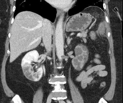
DOI: https://doi.org/10.21980/J8804KOn the coronal sections of computed tomography (CT), bilateral renal infarctions (blue arrows) and several splenic infarctions (green arrows) are noted. Of particular interest, part of the clot totally occluding the left renal artery visibly extends into the aorta (red arrow). The vascular reconstruction image is remarkable for the absent left kidney, the unusual contour of the right kidney and the abnormal splenic blush.
Saddle Pulmonary Embolus
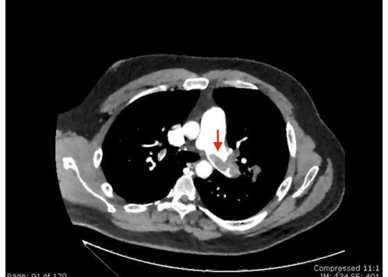
DOI: https://doi.org/10.21980/J8N63PAn electrocardiogram (ECG) showed evidence of right heart strain with an incomplete right bundle branch block, S1Q3T3 (see red arrow [S1], blue arrow [Q3], and black arrow [T3]), and ST-segment elevation in the septal leads (green arrows). Bedside echocardiography showed a dilated right ventricle with ventricular wall akinesis (red arrow) sparing the apex (purple arrow), which is known as McConnell’s Sign. It also showed a mobile hyperechoic mass (yellow arrow). These ultrasound findings were concerning for pulmonary embolism (PE), so computed tomography (CT) angiogram of the chest was ordered and confirmed massive bilateral obstructive filling defects (red arrows) consistent with saddle pulmonary embolism. Additionally, noted is flattening of the interventricular septum (blue arrow) consistent with right heart strain. Laboratory studies were notable for a troponin-I of 0.29 ng/mL, a B-type natriuretic peptide of 792.3 pg/mL, lactic acid of 5.30 mmol/L, and a creatinine of 2.0 mg/dL, consistent with end organ dysfunction. All other lab work was within normal limits.
Sigmoid Diverticulitis Complicated by Colovesical Fistula Presenting with Pneumaturia
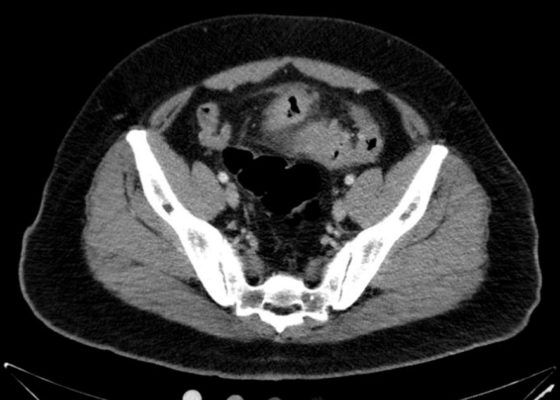
DOI: https://doi.org/10.21980/J80G9TA CT scan of his abdomen/pelvis shows acute sigmoid colonic diverticulitis with adjacent extraluminal collection containing gas (axial view, white arrow) consistent with perforation, along with abutment of the urinary bladder with intraluminal bladder gas (sagittal and coronal views, white arrowheads) suggesting colovesical fistula.
Beware the Devastating Outcome of a Common Procedure
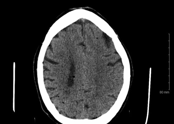
DOI: https://doi.org/10.21980/J8T336Non-contrast head computed tomography (CT) demonstrates multifocal bilateral hypodense lesions (white arrows) representing air emboli. Note the lesions are located in the intra-axial distribution which indicates an underlying vascular origin.
Ovarian Teratoma
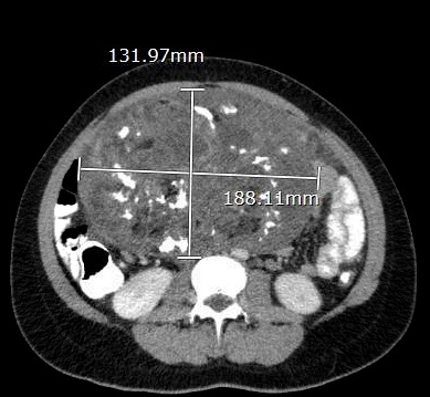
DOI: https://doi.org/10.21980/J8934XThe CT scan with oral contrast in the emergency department revealed a large heterogeneous abdominopelvic mass measuring 13.2 x 18.8 x 23.1 cm (see white lines), suggestive of an ovarian teratoma from the right ovary. This mass included fat, fluid, calcifications (see yellow arrows), and enhancing soft tissue components. The teratoma resulted in mass effect upon large and small bowel loops (see blue highlighted areas), inferior vena cava (IVC), distal aorta (see red highlighted area) and right common iliac artery. A small volume of ascites was also observed. There was no evidence of bowel obstruction, vascular occlusion or other significant emergent finding. Additionally, transabdominal and transvaginal ultrasound images were obtained. The transabdominal image visualized the abdominopelvic mass (see four yellow stars). The transvaginal image visualized a cross section of the teratoma (see four red stars) in relation to the bladder (see four blue stars).
Intramural Hematoma with Type B Aortic Dissection
DOI: https://doi.org/10.21980/J81M03Computed tomography angiography of the chest and abdomen revealed a 9.5 cm thoracoabdominal aneurysm (red outline) with intramural hematoma (yellow shading) and large left pleural effusion versus hemothorax with old blood (blue shading).
Open Globe with Intraocular Foreign Body
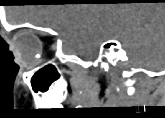
DOI: https://doi.org/10.21980/J8S348On physical exam, his extraocular movements were intact. The right anterior chamber appeared cloudy, particularly nasal to the pupil. The conjunctiva of the right eye was injected. The right pupil was 3 mm and sluggishly reactive and appeared slightly irregular (see yellow arrow). Of note, the right eye also had a 1 mm hypopyon, indicating inflammation of the anterior chamber, which was visible on slit lamp examination (not pictured). There was no fluorescein uptake or Seidel sign. His visual acuity was 20/60 OD (right eye) and 20/20 OS (left eye).
Endocarditis
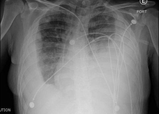
DOI: https://doi.org/10.21980/J8JP73Upright frontal radiograph of the chest demonstrated large pleural effusion on the left and moderate pleural effusion on the right as shown by the visible menisci on both sides (red arrows) with diffuse bilateral nodular densities (yellow dotted lines), consistent with septic pulmonary emboli. Computed tomography (CT) of the chest demonstrated multiple scattered lung nodules bilaterally containing internal foci of air cavitation (green dotted lines).

