Visual EM
Case Report of Distal Radioulnar Joint and Posterior Elbow Dislocation
DOI: https://doi.org/10.21980/J89S6KRadiographs of the left elbow and wrist were obtained. Left elbow radiographs showed simple posterolateral dislocation of the olecranon (red) without fracture of the olecranon (red) or trochlea (blue). Left wrist lateral radiographs demonstrated DRUJ dislocation with dorsal displacement of the distal ulna (green) without fracture or widening of the radioulnar joint (purple). Post-reduction radiographs demonstrated appropriate alignment of the elbow with the trochlea seated in the olecranon and improved alignment of the DRUJ.
Bladder Diverticulum – A Case Report
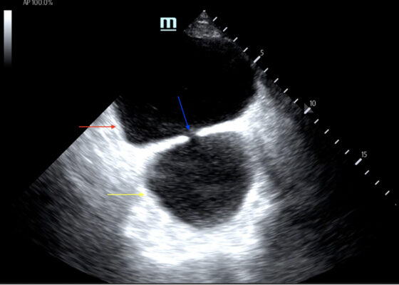
DOI: https://doi.org/10.21980/J8635COn examination, the patient was alert and oriented but in mild distress. Suprapubic fullness was noted upon abdominal palpation. Point of care ultrasound of the bladder showed two enlarged “bladders” with a central communication. Bedside total bladder volume was measured to be 1288 cm3 (the top “bladder” was measured to be 1011 cm3, while the bottom “diverticulum” was measured to be 277 cm3) by ultrasound.
The POCUS stills of the patient’s bladder demonstrated the bladder (red arrow) and bladder diverticulum (yellow arrow) with a central communication (blue arrow) in the transverse and sagittal views.
A Case Report of Ogilvie’s Syndrome in a 58-year-old Quadriplegic
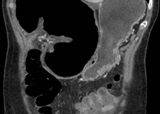
DOI: https://doi.org/10.21980/J82922Plain radiograph of the patient's abdomen revealed a gaseous distention of the colon. This is demonstrated as noted in the abdominal x-ray as gaseous distention, most notably in the large bowel (arrows) including the rectal region (large circle). Follow up computed tomography (CT) scan affirmed severe pancolonic gaseous distention measuring up to 11.2 cm, compatible with colonic pseudo-obstruction as noted by the large red arrows. No anatomical lesion or mechanical obstruction was observed, as well as no evidence of malignancy or other acute process.
Cecal Volvulus Diagnosed with a Whirl Sign: A Case Report
DOI: https://doi.org/10.21980/J8XM05The CT image demonstrates a “whirl sign” (red arrow) which is indicative of a volvulus. This image occurs when bowel, mesentery and vasculature rotate around a transition point causing an image similar to a hurricane on a weather map. When seen on a CT scan, a whirl sign suggests a high likelihood of either a closed loop bowel obstruction or volvulus in the cecum, sigmoid or midgut. In any of the cases, seeing a whirl sign strongly increases the need for emergent surgical management.
Paroxysmal Ventricular Standstill—A Case Report of all Ps and no QRS in Ventricular Asystole
DOI: https://doi.org/10.21980/J8SS79In route, it was proposed that this patient was suffering from a dysrhythmia due to the transient episodes of syncope with lack of ventricular activity on telemetry. Upon close examination of the rhythm strips as well as the ECG, P waves can be visualized without any accompanying QRS complexes lasting multiple seconds (ED ECG blue arrows). Additionally, the rhythm has an intrinsic rate of 100 beats per minute and has a consistent morphology with no evidence of ventricular activity due to the lack of QRS complexes. As a result, the rhythm likely originates in the atria with no passage of impulses into the ventricles through the atrioventricular (AV) node versus an accelerated ventricular rhythm where QRS complexes would be seen.8 These rhythm strips demonstrate an example of VS. There is preserved native atrial automaticity, with an intact sinoatrial (SA) node, with a complete lack of ventricular electrical activity
Severe Hyperkalemia
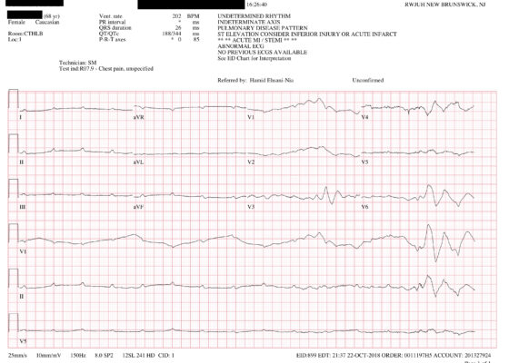
DOI: https://doi.org/10.21980/J8KH1DThe initial ECG obtained upon arrival shows what is commonly referred to as a sine wave pattern. This patient does have a biventricular pacemaker which would ordinarily create a wide QRS complex mimicking an intraventricular conduction delay. However, the QRS complex here is exceptionally wide, in excess of 400 milliseconds (normal: less than 120 milliseconds). As the QRS widens, alongside other deflections present on the ECG, it morphologically mimics a mathematical sine wave.
Is an X-ray a Useful Test for Esophageal Food Boluses? A Case Report
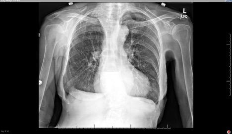
DOI: https://doi.org/10.21980/J8Q639The plain film radiograph of the chest demonstrated a fluid level (yellow arrow) in the distal esophagus with dilation of the esophagus proximal to that point (blue line). Neither of these findings were present on the previous visit.
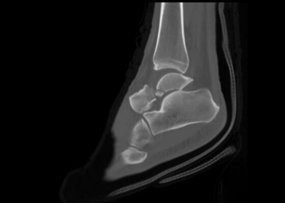
Case Report: Talar Neck Fracture
DOI: https://doi.org/10.21980/J8FP75ABSTRACT: This report demonstrates a case of a severe talar neck fracture. Although rare, talar neck fractures have a high potential for morbidity. Typically caused by a high energy injury, this patient’s mechanism of injury was relatively minor, and presentation was not immediately concerning for such a severe fracture. Initial x-rays provided a gross demonstration of the fracture, but a


