
JETem
  • Home
  • About
    • Aim and Scope
    • Our Team
    • Editorial Board
    • FAQ
  • Issues
    • Current Issue
    • Ahead of Print
    • Past Issues
  • Visual EM
    • Latest Visual EM
    • Search Visual EM
    • Thumbnail Library
  • For Authors
    • Instructions for Authors
    • Submit to JETem
    • Photo Consent
    • Policies
      • Peer Review Policy
      • Copyright Policy
      • Editorial Policy, Ethics and Responsibilities
      • Conflicts of Interest & Informed Consent
      • Open Access Policy
      • Acceptance Fee
  • For Reviewers
    • Instructions for JETem Reviewers
    • Interested in Being a JETem Reviewer?
  • Topic
    • Abdominal/Gastroenterology
    • Administration
    • Board Review
    • Cardiology/Vascular
    • Certifying Exam Cases
      • Clinical Decision-Making
      • Prioritization
      • Communication
      • Procedure & Ultrasound
    • Clinical Informatics, Telehealth and Technology
    • Dermatology
    • EMS
    • Endocrine
    • ENT
    • Faculty Development
    • Genitourinary
    • Geriatrics
    • Hematology/Oncology
    • Infectious Disease
    • Miscellaneous (stats, etc)
    • Neurology
    • Ob/Gyn
    • Ophthalmology
    • Orthopedics
    • Pediatrics
    • Pharmacology
    • Procedures
    • Psychiatry
    • Renal/Electrolytes/Nutrition
    • Respiratory
    • Social Determinants of Health
    • Toxicology
    • Trauma
    • Ultrasound
    • Urology
    • Wellness
    • Wilderness
  • Modality
    • Certifying Exam Practice Cases
      • Clinical Care Cases
      • Communication & Procedural Cases
      • Old School Oral Board Cases
    • Curricula
    • Innovations
    • Lectures
    • Podcasts
    • Simulation
    • Small Group Learning
    • Team Based Learning (TBL)
    • Visual EM
  • Contact Us

Visual EM

Creative Commons images

Elderly female with acute abdominal pain presenting with Superior Mesenteric Artery Thrombus

Sassan Ghassemzadeh, MD*

DOI: https://doi.org/10.21980/J82W52 Issue 3:2[mrp_rating_result]
Computed tomography (CT) angiogram of the abdomen and pelvis revealed a superior mesenteric artery (SMA) thrombosis 5 cm from the origin off of the abdominal aorta. As seen in the sagittal view, there does not appear to be any contrast 5 cm past the origin of the SMA. On the axial views, you can trace the SMA until the point that there is no longer any contrast visible which indicates the start of the thrombus.  The SMA does not appear to be reconstituted. There was normal flow to the celiac artery. (See annotated images).
Abdominal/GastroenterologyCardiology/VascularVisual EM
Creative Commons images

Biceps Tendon Rupture

Daniel M Polvino, MD* and Grant Wei, MD*

DOI: https://doi.org/10.21980/J8RP8B Issue 3:1[mrp_rating_result]
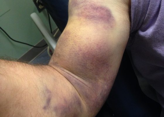
Physical exam was significant for ecchymosis and mild swelling of the right bicep. When the right arm was flexed at the elbow, a prominent mass was visible and palpable over the right bicep. Right upper extremity strength was 4/5 with flexion at the elbow.
OrthopedicsVisual EM
Creative Commons images

Hutchinson’s Sign

Lawrence Lau, MD*, Hannah Mirzakhani, BA^ and Nicholas Governatori, MD*

DOI: https://doi.org/10.21980/J8N040 Issue 3:1[mrp_rating_result]
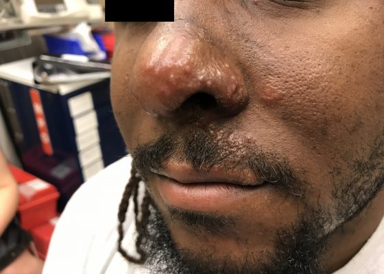
The unilateral distribution of vesicular lesions over the patient's left naris, cheek, and upper lip are consistent with Herpes zoster reactivation with Hutchinson's sign.  Hutchinson's sign is a herpes zoster vesicle present on the tip or side of the nose.1 It reflects zoster involvement of the 1st branch of the trigeminal nerve, and is concerning for herpes zoster ophthalmicus.1 Herpes zoster vesicles may present as papular lesions or macular vesicles on an erythematous base.2,3 Emergent diagnosis must be made to prevent long-term visual sequelae.4
Infectious DiseaseDermatologyVisual EM
Creative Commons images

Viridans streptococci Intracranial Abscess Masquerading as Metastatic Disease

Brandon Ruderman, MD*, Traci Thoureen, MD* and Joshua Broder, MD*

DOI: https://doi.org/10.21980/J8CH05 Issue 3:1[mrp_rating_result]
A non-contrast CT (Figure 1) revealed a large hypoattenuating left parietal lesion. When the CT was enhanced with intravenous contrast (Figure 2), the same lesion showed peripheral rim enhancement, suggestive of a brain abscess.
Infectious DiseaseNeurologyVisual EM
Creative Commons images

A Sublingual Mass in a 5-Year-Old Male

Chris A Rees MD, MPH*, Andrea T Cruz, MD, MPH^ and Corrie E Chumpitazi, MD, MS^

DOI: https://doi.org/10.21980/J8H622Issue 3:1[mrp_rating_result]
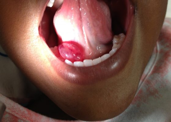
Oral ranula. On oral inspection he had a 3 x 1.5 cm, soft, non-tender, mobile, cystic, sublingual mass on the right aspect of the floor of his mouth that did not move with swallowing (Image 1). There was mild associated submandibular swelling on the right side of his face.
ENTVisual EM
Creative Commons images

Pneumomediastinum After Cervical Stab Wound

Chad Correa, BS* and Emily Ma, MD^

DOI: https://doi.org/10.21980/J87P79 Issue 3:1[mrp_rating_result]
Anteroposterior (AP) chest X-ray showed subcutaneous emphysema of the neck, surrounding the trachea (red arrows), right side greater than left, and a streak of gas adjacent to the aortic arch (white arrow). Computed tomography angiogram (CTA) of the neck showed air outside of the trachea, positive for pneumomediastinum (blue arrows). 
TraumaRespiratoryVisual EM
Creative Commons images

A Case of Acute Cholecystitis

Chad Correa, BS* and Lindsey Spiegelman, MD^

DOI: https://doi.org/10.21980/J8405QIssue 3:1[mrp_rating_result]
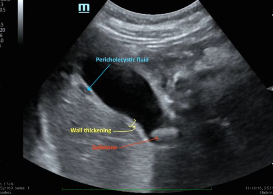
Bedside point-of-care ultrasound revealed a distended gallbladder, thickened gallbladder wall, pericholecystic fluid, and a stone in the neck of the gallbladder indicative of acute cholecystitis.
Abdominal/GastroenterologyVisual EM
Creative Commons images

Computed Tomography and Ultrasound Diagnosis of Spontaneous Subcapsular Renal Hematoma

Samer Assaf, MD*, Thalia Nguyen* and Alisa Wray, MD*

DOI: https://doi.org/10.21980/J8062DIssue 3:1[mrp_rating_result]
Bedside ultrasound was performed and demonstrated a hypoechoic area within the left kidney (images not shown). The non-contrast computed tomography (CT) of the abdomen and pelvis shows a significantly enlarged left kidney and a region of high-attenuation encapsulating the left kidney, concerning for acute hemorrhage.
Renal/ElectrolytesVisual EM
«‹34›»
Page 34 of 45

JETem is an online, open access, peer-reviewed, journal-repository for EM educators. We are PMC Indexed.

Most Viewed

  • Simple Auricular Hematoma (“Cauliflower Ear”) Task Trainer
  • The Three Breath Method: Teaching Coping Mechanisms for Processing Patient Death
  • Clinical Decision Making: Acute Ischemic Stroke with Large Vessel Occlusion
  • PRIMER: Premedical Introduction to Mentored Emergency Research: Connecting Undergraduate Assistants and Emergency Medicine Residents in a Community Hospital Setting
  • Dangerous Dysrhythmias in the ED: An Interactive EKG Workshop for Emergency Medicine Clerkship Learners

Visit Our Collaborators


Creative Commons Licence
This work is licensed under a Creative Commons Attribution 4.0 International License.

About

Education

Learners should benefit from active learning. JETem accepts submissions of team-based learning, small group learning, simulation, podcasts, lectures, innovations, curricula, question sets, and visualEM.

Scholarship

We believe educators should advance through the scholarship of their educational work. JETem gives educators the opportunity to publish scholarly academic work so that it may be widely distributed, thereby increasing the significance of their results.

Links

  • Home
  • Aim and Scope
  • Current Issue
  • For Reviewers
  • Instructions for Authors
  • Contact Us

Newsletter

Sign up to receive updates from JETem regarding newly published issues and findings.

Copyright Creative Commons Attribution 4.0 International