Ultrasound
The Role of Chest X-Ray and Bedside Ultrasound in Diagnosing Pulmonary Bleb versus Pneumothorax
DOI: https://doi.org/10.21980/J8MP7QThe patient was evaluated with bedside ultrasound for concern of possible pneumothorax. Imaging of the left lung with M-mode demonstrated a “sea shore” sign showing a wavy pattern below the pleural line caused by lung sliding as well as “comet tail” artifact caused by from the deep pleura. However, there was no lung sliding on the right shown by a lack of “comet tail” artifact and a “bar code” sign where M-mode shows straight lines throughout the image, this is caused by lack of motion below the pleura. This lack of lung sliding is consistent with possible pneumothorax or bleb.
A two-view chest X-ray (CXR) revealed absent lung parenchyma in the right lung similar to a large pneumothorax (see red outline). Electronic medical record chart review revealed previous CXRs with similar findings. This patient was determined to have an acute COPD exacerbation with chronic blebs, but no pneumothorax.
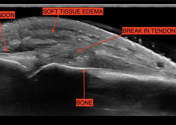
Fight Bite with Tendon Laceration
DOI: https://doi.org/10.21980/J8MP7QThe video shows a water bath ultrasound of the right 4th digit, demonstrating soft tissue swelling with a hypoechoic region along the tendon consistent with edema and tendon disruption (see video and annotated still image).
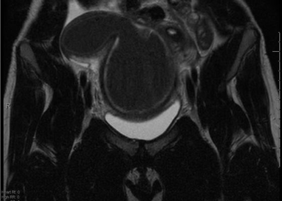
A Rare Cause of Pelvic Pain in a Teenage Girl
DOI: https://doi.org/10.21980/J87D0WDue to pain out of proportion to her exam, an ultrasound of her pelvis was obtained and showed a blood-filled distended uterus, or hematometrocolpos (white arrow), with a 4.9 cm right ovarian cyst (blue arrow). A pelvic magnetic resonance imaging (MRI) then revealed an obstructed right hemi-vagina, normal left uterus and vagina and ipsilateral renal agenesis (red arrow) with normal left kidney (double arrow) consistent with obstructed hemivagina, ipsilateral renal agenesis (OHVIRA) syndrome. The patient underwent surgical repair with complete resolution of symptoms.
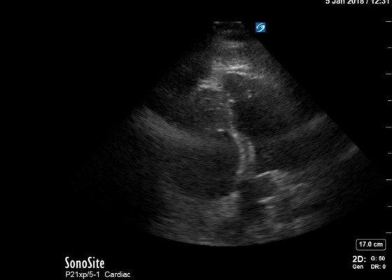
Right Ventricular Dilation in Patient With Submassive Pulmonary Embolism
DOI: https://doi.org/10.21980/J82P84Bedside echocardiography four chamber view revealed enlarged right ventricular (RV) to left ventricular (LV) ratio (greater than 1) on apical four-chamber view (see red and blue outlines respectively). The right atrium is not clearly delineated in this image and therefore is not outlined. One can also rule out a large pericardial effusion as the cause of her dyspnea, since there is no large hypoechoic collection surrounding the heart on either four- chamber view or parasternal long view.
Point of Care Ultrasound Illustrating Small Bowel Obstruction
DOI: https://doi.org/10.21980/J8T637POCUS of the small bowel illustrated significantly dilated loops of bowel (white line), thickened bowel wall (white arrow) and to-and-fro peristalsis, consistent with small bowel obstruction.
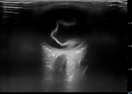
Retinal Detachment
DOI: https://doi.org/10.21980/J8204QBedside ocular ultrasound revealed a serpentine, hyperechoic membrane that appeared tethered to the optic disc posteriorly with hyperechoic material underneath. These findings are consistent with retinal detachment (RD) and associated retinal hemorrhage.
Intussusception
DOI: https://doi.org/10.21980/J8SH0WA segment of bowel within the right abdomen that measured approximately 1.6 x 1.5 cm transaxially. It demonstrated a hypoechoic edematous outer loop of bowel (blue arrow) and hyperechoic compressed loop of bowel telescoping within (red star), this is known as the "target sign."
Evaluation of Snake Bites with Bedside Ultrasonography
DOI: https://doi.org/10.21980/J84S7DHistory of present illness: While watering his lawn, a 36-year-old man felt two sharp bites to his bilateral ankles. He reports that he then saw a light brown, 2-foot snake slither away from him. He came to the emergency department because of pain and swelling in his ankles and inability to bear weight. Physical examination revealed bilateral ankle swelling and


