Bedside Echocardiography for Rapid Diagnosis of Malignant Cardiac Tamponade
History of present illness:
A 47-year-old female with metastatic breast cancer presented to the Emergency Department with chest pain and shortness of breath. She was hypotensive and her EKG showed sinus tachycardia with low voltage. A bedside ultrasound was performed that detected a pericardial effusion and evidence of cardiac tamponade. The patient’s vitals improved with a fluid bolus and she went emergently to the cardiac catheterization lab for fluoroscopy and echocardiography guided pericardiocentesis. A total of 770 mL of fluid was removed from her pericardial space.
Significant findings:
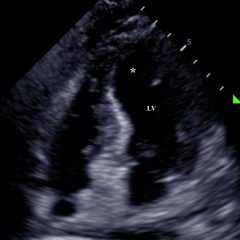
The video shows a subxiphoid view of the heart with evidence of a large pericardial effusion with tamponade – note the anechoic stripe in the pericardial sac (see red arrow). This video demonstrates paradoxical right ventricular collapse during diastole and right atrial collapse during systole which is indicative of tamponade.1,2
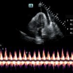
The still image is from the same patient and shows sonographic pulsus paradoxus. This is an apical 4 chamber view of the heart with the sampling gate of the pulsed wave doppler placed over the mitral valve. The Vpeak max and Vpeak min are indicated. If there is more than a 25% difference with inspiration between these 2 values, this is highly suggestive of tamponade.1 In this case, there is a 32.4% difference between the Vpeak max 69.55 cm/s and Vpeak min 46.99 cm/s.
Discussion:
Cardiac tamponade is distinguished from pericardial effusion by right ventricular compression/collapse and hemodynamic instability. Findings can include hypotension, tachycardia, distant heart sounds, and jugular venous distension.3,4 One might also see a plethoric IVC without respiratory variation indicative of elevated right atrial pressures.1 Detection of right ventricular collapse for cardiac tamponade has sensitivities ranging from 48%-100% and specificities ranging from 33%-100%.5 A larger effusion is more likely to lead to cardiac tamponade. However, size of the effusion is less important than the rate of the fluid accumulation.6 For example, cancer patients may come in with large but chronic malignant pericardial effusions and be completely asymptomatic while young, healthy patients with only a small amount of hemopericardium may be on the brink of complete cardiovascular collapse. Utilizing bedside echocardiography allows for prompt diagnosis of a potentially rapidly fatal condition.
Topics:
Echocardiography, ultrasound, cardiac, cardiology, tamponade, effusion, pericardial.
References:
- Ginghina C, Beladan CC, Iancu M, Calin A, BA Popescu. Respiratory maneuvers in echocardiography: a review of clinical applications. Cardiovasc Ultrasound. 2009;7(42):1-13. doi: 10.1186/1476-7120-7-42
- Singh S, Wann LS, Schuchard GH, Klopfenstein HS, Leimgruber PP, Keelan MH Jr, et al. Right ventricular and right atrial collapse in patients with cardiac tamponade – a combined echocardiographic and hemodynamic study. Circulation. 1984;70(6):966-971.
- Jehangir W, Osman M. Electrical alternans with pericardial tamponade. N Engl J Med. 2015;373(8):e10. doi: 10.1056/NEJMicm1408805
- Carmody K, Asaly M, Blackstock U. Point of care echocardiography in an acute thoracic dissection with tamponade in a young man with chest pain, tachycardia, and fever. J Emerg Med. 2016;51(5):e123-e126. doi: 10.1016/j.jemermed.2016.06.046
- Guntheroth WG. Sensitivity and specificity of echocardiographic evidence of tamponade: implication for ventricular interdependence and pulsus paradoxus. Pediatr Cardiol. 2007;28(5):358-62. doi: 10.1007/s00246-005-0807-9
- Butala A, Chaudhari S, Sacerdote A. Cardiac tamponade as a presenting manifestation of severe hypothyroidism. BMJ Case Rep. 2013. doi: 10.1136/bcr-12-2011-5281


