• Registration
  • Login
JETem
  • Home
  • About
    • Aim and Scope
    • Our Team
    • Editorial Board
    • FAQ
  • Issues
    • Current Issue
    • Ahead of Print
    • Past Issues
  • Visual EM
    • Latest Visual EM
    • Search Visual EM
    • Thumbnail Library
  • For Authors
    • Instructions for Authors
    • Submit to JETem
    • Photo Consent
    • Policies
      • Peer Review Policy
      • Copyright Policy
      • Editorial Policy, Ethics and Responsibilities
      • Conflicts of Interest & Informed Consent
      • Open Access Policy
  • For Reviewers
    • Instructions for JETem Reviewers
    • Interested in Being a JETem Reviewer?
  • Topic
    • Abdominal / Gastroenterology
    • Administration
    • Board Review
    • Cardiology / Vascular
    • Clinical Informatics, Telehealth and Technology
    • Dermatology
    • EMS
    • Endocrine
    • ENT
    • Faculty Development
    • Genitourinary
    • Geriatrics
    • Hematology / Oncology
    • Infectious Disease
    • Miscellaneous
    • Neurology
    • Ob / Gyn
    • Ophthalmology
    • Orthopedics
    • Pediatrics
    • Pharmacology
    • Procedures
    • Psychiatry
    • Renal / Electrolytes
    • Respiratory
    • Social Determinants of Health
    • Toxicology
    • Trauma
    • Ultrasound
    • Urology
    • Wellness
    • Wilderness
  • Modality
    • Curricula
    • Innovations
    • Lectures
    • Oral Boards
      • Structured Interview
      • Communication Case
    • Podcasts
    • Simulation
    • Small Group Learning
    • Team Based Learning
    • Visual EM
  • Contact Us

Issue 3:4

Creative Commons images

Fournier Gangrene

Joseph Zakaria, BS*, Steve Bunch, MD* and Victor Cisneros, MD, MPH*

DOI: https://doi.org/10.21980/J89626Issue 3:4 No ratings yet.
The computed tomography (CT) of the abdomen and pelvis revealed significant subcutaneous gas tracking along the perineum and right gluteal region (orange outline) into the scrotum with associated scrotal edema (yellow arrow) and subcutaneous inflammatory fat stranding of 0.92 cm (red arrow) consistent with Fournier’s gangrene.  There is early fluid loculation along the right medial gluteal cleft of 5.85 cm (green arrow) without a sizeable drainable abscess seen.
Infectious DiseaseVisual EM
Creative Commons images

Foreign Body in Maxillary Sinus: A Rare Case of Chronic Rhinosinusitis

Richard Barnett, DO* and Meigra Chin, MD*

DOI: https://doi.org/10.21980/J85H09Issue 3:4 No ratings yet.
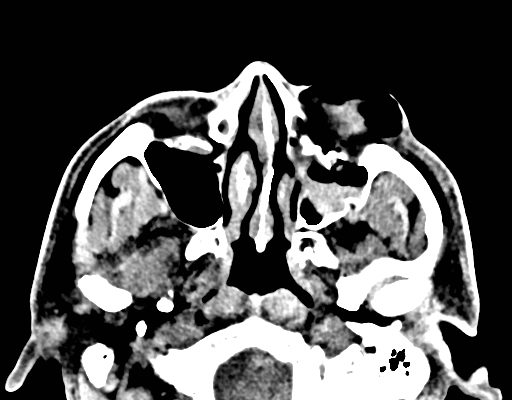
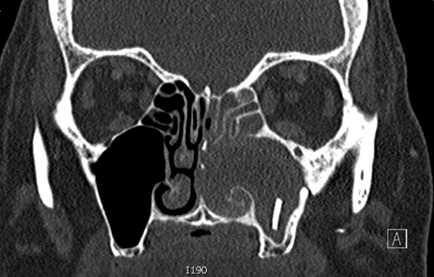
Computed tomography (CT) sinus with contrast demonstrated complete opacification of left paranasal sinuses and nasal cavity, and a linear radiopacity within the left maxillary sinus consistent with a foreign body.  There were additional left facial subcutaneous radiopaque opacities.
ENTVisual EM
Creative Commons images

Brief Review of Intussusception Diagnosis and Management

Claire Thomas, MD*, Wirachin Hoonpongsimanont, MD*, Sara Paradise, MD* and Mohammad Helmy, MD^

DOI: https://doi.org/10.21980/J81P7FIssue 3:4 No ratings yet.
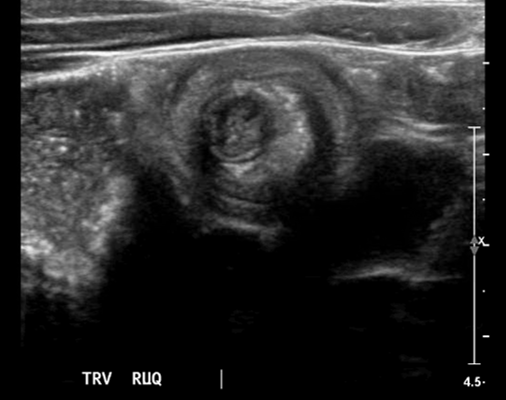
The patient’s abdominal ultrasound revealed intussusception in the right upper abdominal quadrant. The transverse ultrasound view showed a “doughnut sign” (dashed yellow line), telescoping bowel (yellow arrow), and invaginated hyperechoic mesenteric fat with crescent configuration (dashed orange line).  The sagittal ultrasound view demonstrated the intussusception formed by the outer recipient bowel loop (yellow arrows), invaginated hyperechoic mesenteric fat (orange asterisks), and telescoping bowel centrally (red arrow).
Abdominal/GastroenterologyPediatricsVisual EM
Creative Commons images

Corneal Rust Ring

Joseph Zakaria, BS* and Jonathan Peña, MD*

DOI: https://doi.org/10.21980/J8X067Issue 3:4 No ratings yet.
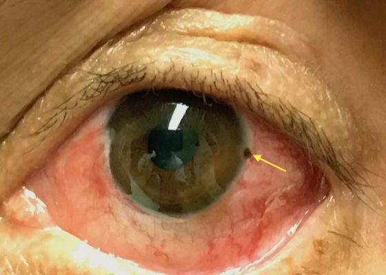
The photograph reveals a limbic metallic foreign body with a surrounding corneal rust ring (arrow) in the three o’clock position of the left cornea. 
OphthalmologyVisual EM
Creative Commons images

Cauliflower Ear Secondary to a Chronic Auricular Hematoma

Nichole Niknafs, DO*

DOI: https://doi.org/10.21980/J8S63XIssue 3:4 No ratings yet.
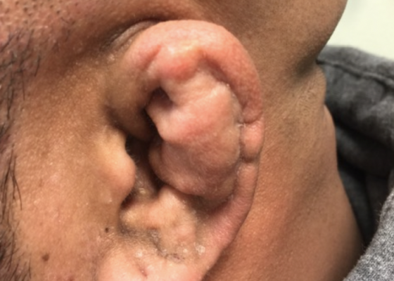
On exam, the patient has a gross deformity to the left pinna that was not painful to touch or fluctuant. Findings and history are consistent with cauliflower ear, secondary to a chronic auricular hematoma.
ENTVisual EM
Creative Commons images

Sialadenitis

Lauren Sylwanowicz, MD* and Andrew Wong, MD, MBA^

DOI: https://doi.org/10.21980/J8NH0NIssue 3:4 No ratings yet.
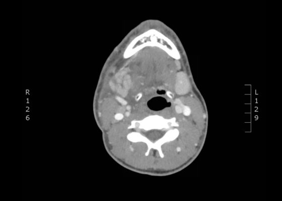
The computed tomography (CT) scan demonstrates prominent enlargement and heterogeneous enhancement of the right submandibular gland (single large arrow) compatible with sialadenitis. There is no evidence of a sialolith or obstruction on the CT. There is associated edema (two small arrows) of the right submandibular space, parapharyngeal space and anterior right neck with partial effacement of the right vallecula and right pyriform sinus.
ENTVisual EM
Creative Commons images

Clinical Evaluation and Management of Pediatric Pericarditis

Sharona Hariri, BA*, Alisa Wray, MD, MAEd^ and Lauren Sylwanowicz, MD^

DOI: https://doi.org/10.21980/J8HP85Issue 3:4 No ratings yet.
An electrocardiogram (ECG) was concerning for ST segment elevation in leads II, III, aVF, and V4, with subtle ST elevations in V5 and V6 (see black arrows).  There is also ST segment depression in aVL (see blue arrows).
Cardiology/VascularVisual EM
Creative Commons images

Point-of-care Ultrasound for the Diagnosis of Ovarian and Fallopian Tube Torsion

Tushank Chadha, BS*, Maili Alexandria Drachman, MD^, Michelle Bui, BS* and Amal Shafi, BS*

DOI: https://doi.org/10.21980/J8D06KIssue 3:4 No ratings yet.
The ultrasound video clip demonstrates a transverse view of the pelvis using the endocavitary probe. The bladder can be seen on the anterior portion of the scan (yellow arrow), while the uterus with an intrauterine pregnancy is visible posteriorly (blue arrow). The thickened appearance of the uterine wall is also indicative of pregnancy. A large, anechoic cystic structure measuring approximately 5 cm is seen in the vicinity of the patient’s left adnexa (pink arrow), which raises concerns for ovarian torsion.
Ob/GynVisual EM
Creative Commons images

Subcutaneous Emphysema After Chest Trauma

Ronald Goubert, BS* and Alisa Wray, MD, MAEd*

DOI: https://doi.org/10.21980/J8864NIssue 3:4 No ratings yet.
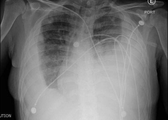
Plain film anteroposterior (AP) radiography of the chest shows left-sided subcutaneous emphysema (red arrow) with overlapping muscle striations of the pectoralis major (green arrow). After chest tube placement (blue arrow), AP chest radiography shows persistent left-sided subcutaneous emphysema (red arrow). CT of the chest shows pneumomediastinum (blue arrow), left apical pneumothorax (pink arrow), and subcutaneous emphysema (red arrow) at the level of T2. At the level of T6, rib fractures can be visualized on the CT (yellow arrow). At the level of T8, left sided pneumothorax is also seen (pink arrow) as the absence of lung tissue on CT.
TraumaRespiratoryVisual EM
Creative Commons images

An Unusual Case of Hematemesis

Amanda Amen, BA*, Jane Xiao, MD^, Julie Parks-Bortel, MD† and Shanna Jones, MD†

DOI: https://doi.org/10.21980/J84H00Issue 3:4 No ratings yet.
The patient’schest X-ray revealed a prominent mediastinum and opacification in the left middle and lower lung fields. The CT showed an aortic aneurysm extending from the thorax to the abdomen with rupture near T7 (blue arrow). It also showed periaortic hemorrhage with active extravasation (green arrow) likely secondary to a penetrating ulcer and bilateral pulmonary opacities concerning for hemothorax (pink arrow).
Cardiology/VascularVisual EM
‹2›
Page 2 of 3

JETem is an online, open access, peer-reviewed, journal-repository for EM educators. We are PMC Indexed.

Most Viewed

  • The Silent Saboteur: Teaching the Clinical Implications of Occult Hypoxemia & Social Determinants of Health via a Pulmonary Embolism Case
  • Diabetic Ketoacidosis and Necrotizing Soft Tissue Infection
  • My Broken Heart
  • Stabilization of Cardiogenic Shock for Critical Care Transport, a Simulation
  • Innovative Ultrasound-Guided Erector Spinae Plane Nerve Block Model for Training Emergency Medicine Physicians

Visit Our Collaborators

Creative Commons Licence
This work is licensed under a Creative Commons Attribution 4.0 International License.

About

Education

Learners should benefit from active learning. JETem accepts submissions of team-based learning, small group learning, simulation, podcasts, lectures, innovations, curricula, question sets, and visualEM.

Scholarship

We believe educators should advance through the scholarship of their educational work. JETem gives educators the opportunity to publish scholarly academic work so that it may be widely distributed, thereby increasing the significance of their results.

Links

  • Home
  • Aim and Scope
  • Current Issue
  • For Reviewers
  • Instructions for Authors
  • Contact Us

Newsletter

Sign up to receive updates from JETem regarding newly published issues and findings.

Copyright Creative Commons Attribution 4.0 International