• Registration
  • Login
JETem
  • Home
  • About
    • Aim and Scope
    • Our Team
    • Editorial Board
    • FAQ
  • Issues
    • Current Issue
    • Ahead of Print
    • Past Issues
  • Visual EM
    • Latest Visual EM
    • Search Visual EM
    • Thumbnail Library
  • For Authors
    • Instructions for Authors
    • Submit to JETem
    • Photo Consent
    • Policies
      • Peer Review Policy
      • Copyright Policy
      • Editorial Policy, Ethics and Responsibilities
      • Conflicts of Interest & Informed Consent
      • Open Access Policy
  • For Reviewers
    • Instructions for JETem Reviewers
    • Interested in Being a JETem Reviewer?
  • Topic
    • Abdominal / Gastroenterology
    • Administration
    • Board Review
    • Cardiology / Vascular
    • Clinical Informatics, Telehealth and Technology
    • Dermatology
    • EMS
    • Endocrine
    • ENT
    • Faculty Development
    • Genitourinary
    • Geriatrics
    • Hematology / Oncology
    • Infectious Disease
    • Miscellaneous
    • Neurology
    • Ob / Gyn
    • Ophthalmology
    • Orthopedics
    • Pediatrics
    • Pharmacology
    • Procedures
    • Psychiatry
    • Renal / Electrolytes
    • Respiratory
    • Social Determinants of Health
    • Toxicology
    • Trauma
    • Ultrasound
    • Urology
    • Wellness
    • Wilderness
  • Modality
    • Curricula
    • Innovations
    • Lectures
    • Oral Boards
      • Structured Interview
      • Communication Case
    • Podcasts
    • Simulation
    • Small Group Learning
    • Team Based Learning
    • Visual EM
  • Contact Us

Issue 3:2

The Use of a Social Media Based Curriculum for Newly Matched Interns Transitioning into Emergency Medicine Residency

Joel C Park, MD*, Miriam Kulkarni, MD* and Mary McLean, MD*

DOI: https://doi.org/10.21980/J8F92GIssue 3:2 No ratings yet.
This curriculum is designed to bridge this gap by fostering an environment in which incoming interns can communicate, collaborate, and practice clinical reasoning with each other and faculty prior to their arrival in residency. The goals and objectives were tailored to the Accreditation Council for Graduate Medical Education (ACGME) level 1 milestones in patient care. 1) Interpret basic ECGs; 2) Demonstrate ability to interpret basic radiographs; 3) Identify common visual diagnoses; 4) Identify common neurological emergencies on head imaging; 5) Recognize basic airway anatomy; 6) Demonstrate successful application of FOAMed resources to clinical cases; 7) Interpret common ultrasound images; 8) Describe common ED procedures; 9) Demonstrate fundamental knowledge of evidence-based medicine and biostatistics 
Miscellaneous (stats, etc)Innovations

Bell’s Palsy

Patrick G Meloy, MD*, Todd A Taylor, MD*, Chris Dudley, MD* and Michelle D Lall, MD*

DOI: https://doi.org/10.21980/J89G9M Issue 3:2 No ratings yet.
At the end of this oral boards session, examinees will: 1) Demonstrate ability to perform a thorough neurologic examination including full cranial nerve exam, National Institutes of Health (NIH) stroke scale assessment, strength and sensation and reflex testing, pronator drift, speech repetition. 2) Differentiate between Bell’s Palsy and acute stroke with facial paralysis. 3) List appropriate laboratory testing for a case of peripheral facial nerve paralysis (basic metabolic panel [BMP]; complete blood count [CBC]; coagulation studies if considering lumbar puncture; human immunodeficiency virus (HIV) test if high-risk by history, or if bilateral; Lyme titer if in endemic area, or if bilateral). 4) Select appropriate treatments (steroids, eye lubricant and patch, PCP referral) for peripheral facial nerve paralysis.
NeurologyOral Boards

High Fidelity In Situ Shoulder Dystocia Simulation

Andrew Pelikan, MD* and Timothy Koboldt, MD*

DOI: https://doi.org/10.21980/J88305D Issue 3:2 No ratings yet.
At the end of this simulation, learners will: 1) Recognize impending delivery and mobilize appropriate resources (ie, both obstetrics [OB] and NICU/pediatrics); 2) Identify risk factors for shoulder dystocia based on history and physical; 3) Recognize shoulder dystocia during delivery; 4) Demonstrate maneuvers to relieve shoulder dystocia; 5) Communicate with team members and nursing staff during resuscitation of a critically ill patient.
Ob/GynSimulation

Fainting Spells

Brittany Guest, DO*, Amir Rouhani, MD* and Steven Lai, MD*

DOI: https://doi.org/10.21980/J8Z91R Issue 3:2 No ratings yet.
ABSTRACT: Audience: The target audience for this simulation is 4th year medical students, emergency medicine residents, pediatric residents, and family medicine residents. Introduction: Brugada syndrome is defined as the combination of specific electrocardiogram (ECG) changes and clinical manifestations of a ventricular arrhythmia, including syncope and sudden cardiac arrest.1 Brugada syndrome is caused by a mutation in the phase-0 cardiac sodium channel. This
Cardiology/VascularSimulation

Guillain-Barrè

Jennifer Yee, DO*, Andrew M King MD* and Geremiha Emerson, MD*

DOI: https://doi.org/10.21980/J8TH06 Issue 3:2 No ratings yet.
At the conclusion of the simulation session, learners will be able to: 1) Recognize the clinical signs and symptoms associated with Guillain-Barré syndrome, including muscle weakness and hyporeflexia. 2) Identify abnormal vital signs secondary to dysautonomia. 3) Discuss evaluation for impending respiratory failure, including bedside pulmonary function testing. 4) Discuss the management of Guillain-Barré, including management of dysautonomia and respiratory failure, as well as definitive management with plasmapheresis versus intravenous immunoglobulin. 5) Appropriately disposition the patient to the intensive care unit. 6) Effectively communicate with team members and nursing staff during resuscitation of a critically ill patient
NeurologySimulation

Sepsis Secondary to an Abdominal Wound Infection

Alisa Wray, MD*

DOI: https://doi.org/10.21980/J8PS60 Issue 3:2 No ratings yet.
At completion of this case learners should be able to: 1) Recognize and differentiate between systemic inflammatory response syndrome, sepsis, severe sepsis, and septic shock. 2) Prepare an appropriate differential diagnosis for a patient with sepsis. 3) Demonstrate appropriate fluid resuscitation and antibiotic therapy for a septic patient. 4) Demonstrate appropriate vasopressor therapy for a septic patient. 5) Understand and apply the Surviving Sepsis Guidelines.
Infectious DiseaseAbdominal/GastroenterologySimulation

Chest Pain with Acute Coronary Syndrome

Alisa Wray, MD*

DOI: https://doi.org/10.21980/J8K04C Issue 3:2 No ratings yet.
At the end of this case learners should be able to prepare a differential for patients with chest pain, provide appropriate medications for a patient with chest pain, recognize an ST segment elevation MI, and appropriately disposition a patient with acute myocardial infarction (AMI).
Cardiology/VascularSimulation
Creative Commons images

An Unusual Case of Pharyngitis: Herpes Zoster of Cranial Nerves 9, 10, C2, C3 Mimicking a Tumor

Jason Cheng, DO*, Gregory Reinhold, DO* and Rahmon Zuckerman, DO*

DOI: https://doi.org/10.21980/J8B05K Issue 3:2 No ratings yet.
On exam, the patient was sitting upright while holding an emesis basin filled with saliva. His voice was noticeably hoarse. Examination of the head and neck revealed vesicular eruptions on the left scalp in the V1 dermatome and on the left mastoid process (Images 1 and 2). Physical exam also shows vesicular eruptions on the left posterior oropharynx that did not cross midline (Image 3).
ENTVisual EM
Creative Commons images

Ramsay Hunt Syndrome

John Costumbrado, MD, MPH* and Ryan Brenchley, MD*

DOI: https://doi.org/10.21980/J85S7QIssue 3:2 No ratings yet.
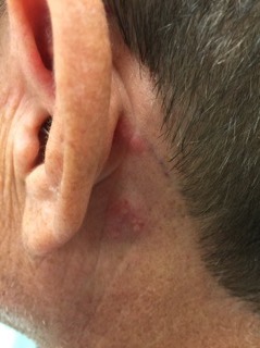
Left-sided cranial nerve VII palsy with flattened forehead creases, inability to keep the left eye open, and drooping of the corner of mouth. Vesicular lesions were found in and posterior to the left ear in a unilateral, dermatomal distribution.
ENTVisual EM
Creative Commons images

Retinal Detachment

Adnan Riaz, MD*, Chirag Shah, MD* and Grant Wei, MD*

DOI: https://doi.org/10.21980/J8204QIssue 3:2 No ratings yet.
Bedside ocular ultrasound revealed a serpentine, hyperechoic membrane that appeared tethered to the optic disc posteriorly with hyperechoic material underneath. These findings are consistent with retinal detachment (RD) and associated retinal hemorrhage.
OphthalmologyUltrasoundVisual EM
1›»
Page 1 of 4

JETem is an online, open access, peer-reviewed, journal-repository for EM educators. We are PMC Indexed.

Most Viewed

  • The Silent Saboteur: Teaching the Clinical Implications of Occult Hypoxemia & Social Determinants of Health via a Pulmonary Embolism Case
  • Diabetic Ketoacidosis and Necrotizing Soft Tissue Infection
  • My Broken Heart
  • Stabilization of Cardiogenic Shock for Critical Care Transport, a Simulation
  • Innovative Ultrasound-Guided Erector Spinae Plane Nerve Block Model for Training Emergency Medicine Physicians

Visit Our Collaborators

Creative Commons Licence
This work is licensed under a Creative Commons Attribution 4.0 International License.

About

Education

Learners should benefit from active learning. JETem accepts submissions of team-based learning, small group learning, simulation, podcasts, lectures, innovations, curricula, question sets, and visualEM.

Scholarship

We believe educators should advance through the scholarship of their educational work. JETem gives educators the opportunity to publish scholarly academic work so that it may be widely distributed, thereby increasing the significance of their results.

Links

  • Home
  • Aim and Scope
  • Current Issue
  • For Reviewers
  • Instructions for Authors
  • Contact Us

Newsletter

Sign up to receive updates from JETem regarding newly published issues and findings.

Copyright Creative Commons Attribution 4.0 International