• Registration
  • Login
JETem
  • Home
  • About
    • Aim and Scope
    • Our Team
    • Editorial Board
    • FAQ
  • Issues
    • Current Issue
    • Ahead of Print
    • Past Issues
  • Visual EM
    • Latest Visual EM
    • Search Visual EM
    • Thumbnail Library
  • For Authors
    • Instructions for Authors
    • Submit to JETem
    • Photo Consent
    • Policies
      • Peer Review Policy
      • Copyright Policy
      • Editorial Policy, Ethics and Responsibilities
      • Conflicts of Interest & Informed Consent
      • Open Access Policy
  • For Reviewers
    • Instructions for JETem Reviewers
    • Interested in Being a JETem Reviewer?
  • Topic
    • Abdominal / Gastroenterology
    • Administration
    • Board Review
    • Cardiology / Vascular
    • Clinical Informatics, Telehealth and Technology
    • Dermatology
    • EMS
    • Endocrine
    • ENT
    • Faculty Development
    • Genitourinary
    • Geriatrics
    • Hematology / Oncology
    • Infectious Disease
    • Miscellaneous
    • Neurology
    • Ob / Gyn
    • Ophthalmology
    • Orthopedics
    • Pediatrics
    • Pharmacology
    • Procedures
    • Psychiatry
    • Renal / Electrolytes
    • Respiratory
    • Social Determinants of Health
    • Toxicology
    • Trauma
    • Ultrasound
    • Urology
    • Wellness
    • Wilderness
  • Modality
    • Curricula
    • Innovations
    • Lectures
    • Oral Boards
      • Structured Interview
      • Communication Case
    • Podcasts
    • Simulation
    • Small Group Learning
    • Team Based Learning
    • Visual EM
  • Contact Us

Issue 2:2

Creative Commons images

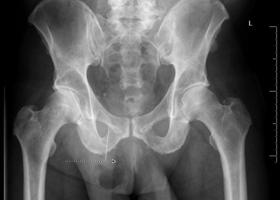
Bowel Perforation complicating an incarcerated inguinal hernia

Adam Sigal, MD* and Jamie Slotkin, DO^

DOI: https://doi.org/10.21980/J8D30BIssue 2:2 No ratings yet.
The AP and lateral pelvis x-rays revealed two sewing needles, 60 mm in length, within the soft tissue over the anterior right lower hemipelvis. In addition, the AP view showed emphysema involving the right hemiscrotum (arrow), concerning for perforated bowel.
Abdominal/GastroenterologyVisual EM
Creative Commons images

Infectious Mononucleosis: Pharyngitis and Morbilliform Rash

Eric McCoy, MD, MPH* and Amal Shafi, BS*

DOI: https://doi.org/10.21980/J88C7H Issue 2:2 No ratings yet.
Her physical exam was significant for bilateral tonsillar exudates, cervical lymphadenopathy, and a morbilliform rash that included the palms (Figure 1-4). Laboratory testing was significant for white blood cell (WBC) count of 16.5 thous/mcl with an elevation in absolute lymphocytes of > 10 thous/mcl. The monospot and EBV (Epstein-Barr virus) panel were positive.
Infectious DiseaseDermatologyENTVisual EM
Creative Commons images

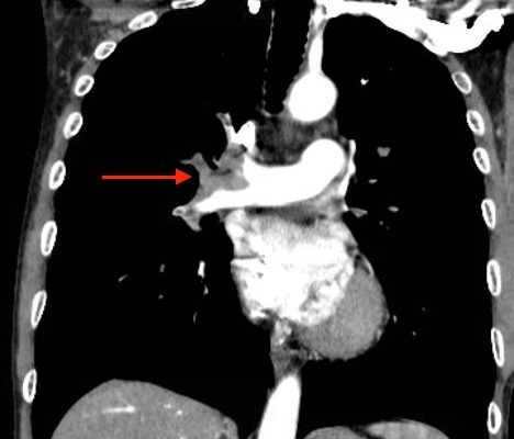
Acute, massive pulmonary embolism with right heart strain and hypoxia requiring emergent tissue plasminogen activator (TPA) infusion

Jonathan Patane, MD* and Wirachin Hoonpongsimanont, MD*

DOI: https://doi.org/10.21980/J84K5K Issue 2:2 No ratings yet.
CT angiogram showed multiple large acute pulmonary emboli, most significantly in the distal right main pulmonary artery (image 1 and 2). Additional pulmonary emboli were noted in the bilateral lobar, segmental, and subsegmental levels of all lobes. There was a peripheral, wedge-shaped consolidation surrounded by groundglass changes in the posterolateral basal right lower lobe that was consistent with a small lung infarction (image 3).
Cardiology/VascularRespiratoryVisual EM
Creative Commons images

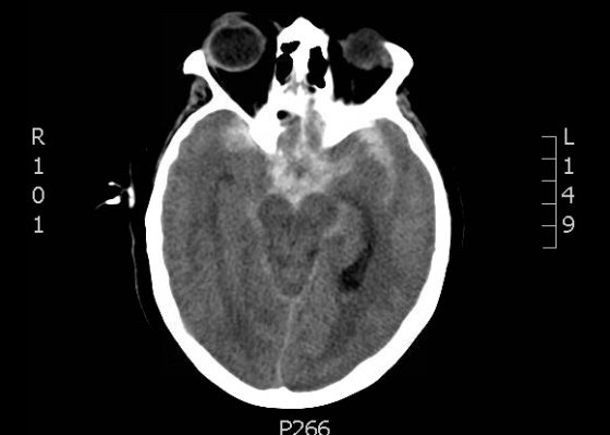
Presentation of Significant Subarachnoid Hemorrhage without Loss of Consciousness

Nicholas Taylor, BA* and Shannon Toohey, MD, MAEd*

DOI: https://doi.org/10.21980/J80W29 Issue 2:2 No ratings yet.
A non-contrast head CT demonstrated extensive subarachnoid hemorrhage occupying both cerebral convexities, the anterior interhemispheric fissure, the sylvian fissures, and the basal cisterns. Later CTA would show an 8 mm by 7 mm by 8mm MCA aneurysm near the M1/M2 junction and two pericallosal artery aneurysms, 7 by 6 mm and 8 by 5 mm respectively.
NeurologyVisual EM
Creative Commons images

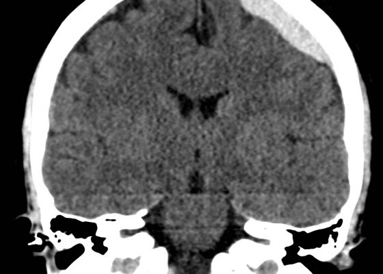
Acute Subdural Hematoma

Ellen Lester, BS*, Jonathan Peña, MD* and Warren Wiechmann, MD, MBA*

DOI: https://doi.org/10.21980/J87C76 Issue 2:2 No ratings yet.
Non-contrast Computed Tomography (CT) of the Head showed a dense extra-axial collection along the left frontal and parietal regions, extending superior to the vertex with mild mass effect, but no midline shift.
NeurologyVisual EM
Creative Commons images

Wellens’ Sign (Wellens’ Syndrome)

Jonathan Patane, MD* and Kim Sokol, MD*

DOI: https://doi.org/10.21980/J8W30P Issue 2:2 No ratings yet.
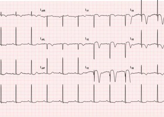
This EKG shows deep, inverted T waves that are most pronounced in V2-V4, and are associated with continued T wave inversions in V5 and V6 and ST segment changes in V1-V3.
Cardiology/VascularVisual EM
Creative Commons images

Herpes Zoster

Hamid Ehsani-Nia, BS, MS* and Robert Rowe, MD^

DOI: https://doi.org/10.21980/J8C301 Issue 2:2 No ratings yet.
The patient was in mild distress, afebrile, with stable vital signs. His physical exam revealed an erythematous, grouped vesicular rash in various stages of progression including erythematous papules, clear vesicles, and pustular vesicles. Few lesions were scabbed over. No signs of crusting or scarring were appreciated. The distribution encompassed the entire left T4 dermatome both posteriorly and anteriorly. No other rashes were appreciated elsewhere on the body.
Infectious DiseaseDermatologyVisual EM
Creative Commons images

Stingray Envenomation

Tanya Dall, MD*

DOI: http://doi.org/10.21980/J86C7W Issue 2:2 No ratings yet.
Physical exam revealed a 3cm laceration to the ulnar side of the dorsum of the left hand with minimal hand swelling. There was no exposed tendon or bone, the hand was neurovascularly intact and had full strength. A small barb was visualized and removed from the injury site. Radiograph of the left hand confirmed that there was no remaining barb (see normal x-ray).
WildernessVisual EM
Creative Commons images

The Lost Guidewire

Ankit Shah, MD*, Adam Sigal, MD* and Kristen Sandel, MD*

DOI: https://doi.org/10.21980/J82P4M Issue 2:2 No ratings yet.
Initial chest radiograph shows a guidewire in the inferior vena cava (IVC), superior vena cava (SVC), and right IJ veins.
ProceduresAdministrationVisual EM
Creative Commons images

A Toddler with Abdominal Pain and Emesis

Saema Said, BS* and Kevin Koenig, MD*

DOI: https://doi.org/10.21980/J8XW2P Issue 2:2 No ratings yet.
In the long axis video, the appendix appears as an enlarged, non-compressible, blind-ending tubular structure (white arrow) with distinct appendiceal wall layers and lack of peristalsis. In the short axis video, the appendix appears as a target sign (yellow arrow) between the abdominal and psoas muscles. The maximal outer diameter (MOD) measures 11.8mm and the appendix wall measures 0.17mm. There is trace adjacent free fluid and echogenic periappendiceal fat. Transverse axis video and image (red arrow) demonstrate that the appendix is not compressible. These findings are consistent with acute appendicitis.
Abdominal/GastroenterologyInfectious DiseasePediatricsUltrasoundVisual EM
‹2
Page 2 of 2

JETem is an online, open access, peer-reviewed, journal-repository for EM educators. We are PMC Indexed.

Most Viewed

  • The Silent Saboteur: Teaching the Clinical Implications of Occult Hypoxemia & Social Determinants of Health via a Pulmonary Embolism Case
  • Diabetic Ketoacidosis and Necrotizing Soft Tissue Infection
  • My Broken Heart
  • Stabilization of Cardiogenic Shock for Critical Care Transport, a Simulation
  • Innovative Ultrasound-Guided Erector Spinae Plane Nerve Block Model for Training Emergency Medicine Physicians

Visit Our Collaborators

Creative Commons Licence
This work is licensed under a Creative Commons Attribution 4.0 International License.

About

Education

Learners should benefit from active learning. JETem accepts submissions of team-based learning, small group learning, simulation, podcasts, lectures, innovations, curricula, question sets, and visualEM.

Scholarship

We believe educators should advance through the scholarship of their educational work. JETem gives educators the opportunity to publish scholarly academic work so that it may be widely distributed, thereby increasing the significance of their results.

Links

  • Home
  • Aim and Scope
  • Current Issue
  • For Reviewers
  • Instructions for Authors
  • Contact Us

Newsletter

Sign up to receive updates from JETem regarding newly published issues and findings.

Copyright Creative Commons Attribution 4.0 International