• Registration
  • Login
JETem
  • Home
  • About
    • Aim and Scope
    • Our Team
    • Editorial Board
    • FAQ
  • Issues
    • Current Issue
    • Ahead of Print
    • Past Issues
  • Visual EM
    • Latest Visual EM
    • Search Visual EM
    • Thumbnail Library
  • For Authors
    • Instructions for Authors
    • Submit to JETem
    • Photo Consent
    • Policies
      • Peer Review Policy
      • Copyright Policy
      • Editorial Policy, Ethics and Responsibilities
      • Conflicts of Interest & Informed Consent
      • Open Access Policy
  • For Reviewers
    • Instructions for JETem Reviewers
    • Interested in Being a JETem Reviewer?
  • Topic
    • Abdominal / Gastroenterology
    • Administration
    • Board Review
    • Cardiology / Vascular
    • Clinical Informatics, Telehealth and Technology
    • Dermatology
    • EMS
    • Endocrine
    • ENT
    • Faculty Development
    • Genitourinary
    • Geriatrics
    • Hematology / Oncology
    • Infectious Disease
    • Miscellaneous
    • Neurology
    • Ob / Gyn
    • Ophthalmology
    • Orthopedics
    • Pediatrics
    • Pharmacology
    • Procedures
    • Psychiatry
    • Renal / Electrolytes
    • Respiratory
    • Social Determinants of Health
    • Toxicology
    • Trauma
    • Ultrasound
    • Urology
    • Wellness
    • Wilderness
  • Modality
    • Curricula
    • Innovations
    • Lectures
    • Oral Boards
      • Structured Interview
      • Communication Case
    • Podcasts
    • Simulation
    • Small Group Learning
    • Team Based Learning
    • Visual EM
  • Contact Us

Issue 2:2

Development of a Head and Neck Regional Anesthesia Task Trainer for Emergency Medicine Learners

Diane L Gorgas, MD*, Sarah Greenberger, MD*, Jillian McGrath, MD*, David P Way, MEd* and Chad Donley, MD^

DOI: https://doi.org/10.21980/J8T595Issue 2:2 No ratings yet.
In participating in the educational session associated with this task trainer, the learner will: 1) Identify landmarks for the following nerve blocks: Infraorbital, Supraorbital (V1), Mental, Periauricular 2) Demonstrate the appropriate technique for anesthetic injection for each of these nerve blocks 3) Map the distribution of regional anesthesia expected from each nerve block 4) Apply the indications and contraindications for each regional nerve block
ProceduresENTInnovations

The Halloween Lateral Canthotomy Model

Nur-Ain Nadir, MD, MHPE*, Ifat Sattar, DO* and Ammar Ahmed, MD*

DOI: https://doi.org/10.21980/J8GW2N Issue 2:2 No ratings yet.
The purpose of the model is to teach residents and students how to perform lateral canthotomy and to achieve competency in their skills.
OphthalmologyInnovations

Emed-Opoly: Echocardiography

Andrew W Phillips, MD, MEd*, Michelle Hunter-Behrend, MD* and Sara Nikravan, MD^

DOI: https://doi.org/10.21980/J8PC77 Issue 2:2 No ratings yet.
By the end of this session, the learner will be able to: 1) Recognize normal and abnormal left heart global function 2) Recognize normal and abnormal right heart global function 3) Recognize pericardial effusions and pericardial tamponade
Cardiology/VascularSmall Group LearningUltrasound

Left Ventricular Assist Devices

Khuansiri Narajeenron, MD, MSc*^, Wirachin Ying Hoonpongsimanot, MD, MS^ and Megan Boysen Osborn, MD, MHPE^

DOI: https://doi.org/10.21980/J8JP4Z Issue 2:2 No ratings yet.
Upon completion of this cTBL module, the learner will be able to: 1) Properly assess LVAD patients’ circulatory status; 2) appropriately resuscitate LVAD patients; 3) identify common LVAD complications; 4) evaluate and appropriately manage patients with LVAD malfunctions.
Cardiology/VascularTeam Based Learning (TBL)

Emergency Medicine Dermatology

Shannon Toohey, MD, MAEd*

DOI: https://doi.org/10.21980/J8DW21 Issue 2:2 No ratings yet.
By the end of this educational session, the learner will: 1) List the six primary types of rash (maculopapular, petechial/purpura, diffuse erythematous, non-erythematous, vesiculo-bullous, and pustular). 2) Be able to accurately describe various types of lesions and rashes with appropriate terminology; 3) Understand the use of the Modified Lynch Algorithm and how it can be used to narrow down the differential diagnosis in patients presenting with rash.
DermatologyTeam Based Learning (TBL)

Pediatric Sepsis Case Scenario

Ilene Claudius, MD*, Sandra Montero, RN*, Madhu Hardasmalani, MD^, Kellyn Pak, RN* and Y. Liza Kearl, MD^

DOI: https://doi.org/10.21980/J8MK5XIssue 2:2 No ratings yet.
Pediatric sepsis is a low-frequency, high impact condition. Nurses and physicians do not see it often, but must recognize and treat children with sepsis efficiently when they present. This makes pediatric sepsis education particularly amenable to simulation scenarios.
PediatricsInfectious DiseaseSimulation

Status Epilepticus in the Emergency Department

Jonathan Lee, BS* and Alisa Wray, MD*

DOI: https://doi.org/10.21980/J8RC7VIssue 2:2 No ratings yet.
At the end of this simulation session, the learner will: 1) Demonstrate the management of status epilepticus 2) Justify when airway intervention is needed for status epilepticus 3) Describe risk factors for status epilepticus 4) Prepare a differential diagnosis for the causes in status epilepticus.
NeurologySimulation
Creative Commons images

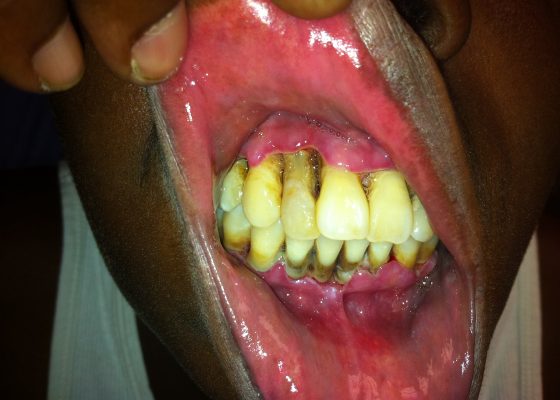
Acute Necrotizing Ulcerative Gingivitis (ANUG)

Nicholas E. Kman, MD* and Vinny P. Kumar, MD*

DOI: https://doi.org/10.21980/J8S88H Issue 2:2 No ratings yet.
Physical examination revealed inflamed gingiva, ulceration, and soft tissue necrosis (Image 1) along with mandibular lymphadenopathy (not shown). Given her symptoms, poor oral care, and her immunocompromised state, she was given a diagnosis of Acute Necrotizing Ulcerative Gingivitis (ANUG) or Vincent’s Angina.
ENTInfectious DiseaseVisual EM
Creative Commons images

Acute Aortic Dissection Presenting Exclusively as Lower Extremity Paresthesias

Ryan Gibney, BS*, Jonathan Patane, MD* and Steven Bunch, MD*

DOI: https://doi.org/10.21980/J8NK57 Issue 2:2 No ratings yet.
Chest x-ray and CT angiogram was performed to evaluate his thoracic and abdominal vasculature. Chest x-ray did not show any significant widening of the mediastinum. The CT angiogram demonstrated an intimal tear along the aortic arch separating a true and false aortic lumen, consistent with an acute aortic dissection. The true lumen (highlighted in blue in images 1-5) can be identified by continuity with an undissected part of the aorta. While the false lumen (highlighted in red in images 1-5) can be identified by its crescent shape and larger cross-sectional area.
Cardiology/VascularVisual EM
Creative Commons images

Galeazzi Fracture

Reid Honda, MD*

DOI: https://doi.org/10.21980/J8HS39 Issue 2:2 No ratings yet.
The X-ray showed an acute comminuted fracture of the distal diaphysis of the radius with disruption of the distal radioulnar joint, consistent with a Galeazzi fracture. The patient was then splinted and taken for operative reduction and internal fixation the following day.
OrthopedicsVisual EM
1›
Page 1 of 2

JETem is an online, open access, peer-reviewed, journal-repository for EM educators. We are PMC Indexed.

Most Viewed

  • The Silent Saboteur: Teaching the Clinical Implications of Occult Hypoxemia & Social Determinants of Health via a Pulmonary Embolism Case
  • Diabetic Ketoacidosis and Necrotizing Soft Tissue Infection
  • My Broken Heart
  • Stabilization of Cardiogenic Shock for Critical Care Transport, a Simulation
  • Innovative Ultrasound-Guided Erector Spinae Plane Nerve Block Model for Training Emergency Medicine Physicians

Visit Our Collaborators

Creative Commons Licence
This work is licensed under a Creative Commons Attribution 4.0 International License.

About

Education

Learners should benefit from active learning. JETem accepts submissions of team-based learning, small group learning, simulation, podcasts, lectures, innovations, curricula, question sets, and visualEM.

Scholarship

We believe educators should advance through the scholarship of their educational work. JETem gives educators the opportunity to publish scholarly academic work so that it may be widely distributed, thereby increasing the significance of their results.

Links

  • Home
  • Aim and Scope
  • Current Issue
  • For Reviewers
  • Instructions for Authors
  • Contact Us

Newsletter

Sign up to receive updates from JETem regarding newly published issues and findings.

Copyright Creative Commons Attribution 4.0 International