Visual EM
Talonavicular Dislocation
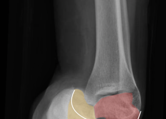
DOI: https://doi.org/10.21980/J8PG91The X-rays were significant for a subtalar dislocation. The calcaneus (red) is laterally displaced with respect to the talar head (orange), and the white lines indicate the normal articular surface. Additionally, there was a talonavicular dislocation, as seen in the fourth image: the talus (green) and navicular bone (purple) overlapping suggests a dislocation. In a normally aligned foot, the boundaries of the two bones create a point of articulation.
Endocarditis
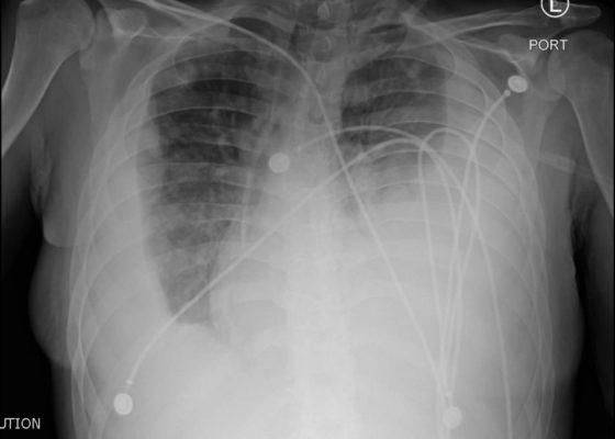
DOI: https://doi.org/10.21980/J8JP73Upright frontal radiograph of the chest demonstrated large pleural effusion on the left and moderate pleural effusion on the right as shown by the visible menisci on both sides (red arrows) with diffuse bilateral nodular densities (yellow dotted lines), consistent with septic pulmonary emboli. Computed tomography (CT) of the chest demonstrated multiple scattered lung nodules bilaterally containing internal foci of air cavitation (green dotted lines).
Facial Fracture Induced Periorbital Emphysema
DOI: https://doi.org/10.21980/J8F05HPhysical exam showed marked left palpebral subcutaneous crepitus, as well as bulbar and palpebral conjunctival bulging. Visual acuity was normal with intact extraocular movements, and normal pupillary exam. Computed tomography (CT) imaging of the face was obtained and revealed multiple displaced fractures involving the left orbital floor and zygomatic arch associated with moderate periorbital and postseptal extraconal gas, resulting in orbital proptosis.
Fournier Gangrene
DOI: https://doi.org/10.21980/J89626The computed tomography (CT) of the abdomen and pelvis revealed significant subcutaneous gas tracking along the perineum and right gluteal region (orange outline) into the scrotum with associated scrotal edema (yellow arrow) and subcutaneous inflammatory fat stranding of 0.92 cm (red arrow) consistent with Fournier’s gangrene. There is early fluid loculation along the right medial gluteal cleft of 5.85 cm (green arrow) without a sizeable drainable abscess seen.
Foreign Body in Maxillary Sinus: A Rare Case of Chronic Rhinosinusitis
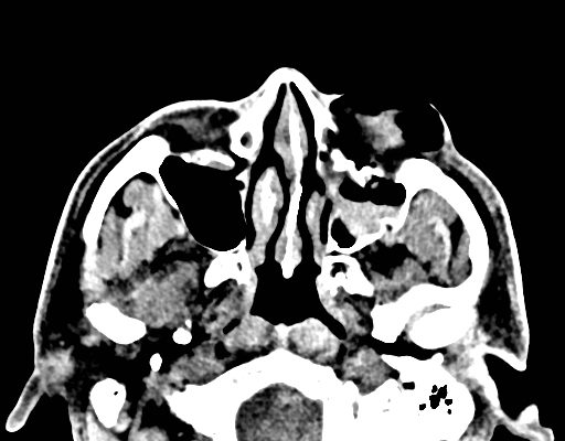
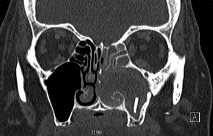
DOI: https://doi.org/10.21980/J85H09Computed tomography (CT) sinus with contrast demonstrated complete opacification of left paranasal sinuses and nasal cavity, and a linear radiopacity within the left maxillary sinus consistent with a foreign body. There were additional left facial subcutaneous radiopaque opacities.
Brief Review of Intussusception Diagnosis and Management
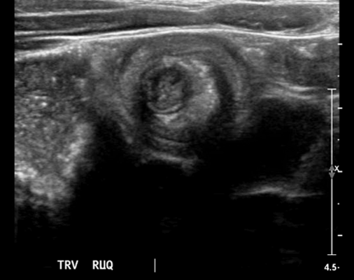
DOI: https://doi.org/10.21980/J81P7FThe patient’s abdominal ultrasound revealed intussusception in the right upper abdominal quadrant. The transverse ultrasound view showed a “doughnut sign” (dashed yellow line), telescoping bowel (yellow arrow), and invaginated hyperechoic mesenteric fat with crescent configuration (dashed orange line). The sagittal ultrasound view demonstrated the intussusception formed by the outer recipient bowel loop (yellow arrows), invaginated hyperechoic mesenteric fat (orange asterisks), and telescoping bowel centrally (red arrow).
Corneal Rust Ring
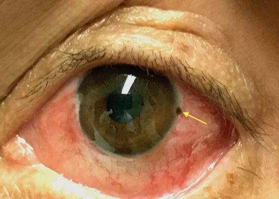
DOI: https://doi.org/10.21980/J8X067The photograph reveals a limbic metallic foreign body with a surrounding corneal rust ring (arrow) in the three o’clock position of the left cornea.
Cauliflower Ear Secondary to a Chronic Auricular Hematoma
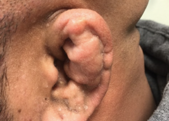
DOI: https://doi.org/10.21980/J8S63XOn exam, the patient has a gross deformity to the left pinna that was not painful to touch or fluctuant. Findings and history are consistent with cauliflower ear, secondary to a chronic auricular hematoma.

