Visual EM
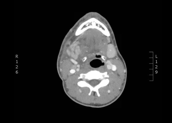
Sialadenitis
DOI: https://doi.org/10.21980/J8NH0NThe computed tomography (CT) scan demonstrates prominent enlargement and heterogeneous enhancement of the right submandibular gland (single large arrow) compatible with sialadenitis. There is no evidence of a sialolith or obstruction on the CT. There is associated edema (two small arrows) of the right submandibular space, parapharyngeal space and anterior right neck with partial effacement of the right vallecula and right pyriform sinus.
Clinical Evaluation and Management of Pediatric Pericarditis
DOI: https://doi.org/10.21980/J8HP85An electrocardiogram (ECG) was concerning for ST segment elevation in leads II, III, aVF, and V4, with subtle ST elevations in V5 and V6 (see black arrows). There is also ST segment depression in aVL (see blue arrows).
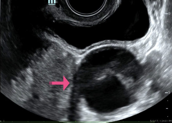
Point-of-care Ultrasound for the Diagnosis of Ovarian and Fallopian Tube Torsion
DOI: https://doi.org/10.21980/J8D06KThe ultrasound video clip demonstrates a transverse view of the pelvis using the endocavitary probe. The bladder can be seen on the anterior portion of the scan (yellow arrow), while the uterus with an intrauterine pregnancy is visible posteriorly (blue arrow). The thickened appearance of the uterine wall is also indicative of pregnancy. A large, anechoic cystic structure measuring approximately 5 cm is seen in the vicinity of the patient’s left adnexa (pink arrow), which raises concerns for ovarian torsion.
Subcutaneous Emphysema After Chest Trauma
DOI: https://doi.org/10.21980/J8864NPlain film anteroposterior (AP) radiography of the chest shows left-sided subcutaneous emphysema (red arrow) with overlapping muscle striations of the pectoralis major (green arrow). After chest tube placement (blue arrow), AP chest radiography shows persistent left-sided subcutaneous emphysema (red arrow). CT of the chest shows pneumomediastinum (blue arrow), left apical pneumothorax (pink arrow), and subcutaneous emphysema (red arrow) at the level of T2. At the level of T6, rib fractures can be visualized on the CT (yellow arrow). At the level of T8, left sided pneumothorax is also seen (pink arrow) as the absence of lung tissue on CT.
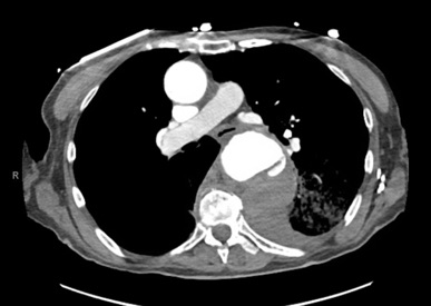
An Unusual Case of Hematemesis
DOI: https://doi.org/10.21980/J84H00The patient’schest X-ray revealed a prominent mediastinum and opacification in the left middle and lower lung fields. The CT showed an aortic aneurysm extending from the thorax to the abdomen with rupture near T7 (blue arrow). It also showed periaortic hemorrhage with active extravasation (green arrow) likely secondary to a penetrating ulcer and bilateral pulmonary opacities concerning for hemothorax (pink arrow).
Extensive Aortic Dissection with Normal Vital Signs
DOI: https://doi.org/10.21980/J80S6SThe patient was found to have a Stanford type A dissection (see yellow arrow) with visible false lumen starting at aortic arch (see green circle). The dissection extended into the descending aorta (see blue circle) as shown by the false lumen (red highlighted area) visible on CT. The radiologist performed a reconstruction of the aorta, which showed that the left kidney was not being perfused, making the kidney not visible on the reconstruction.
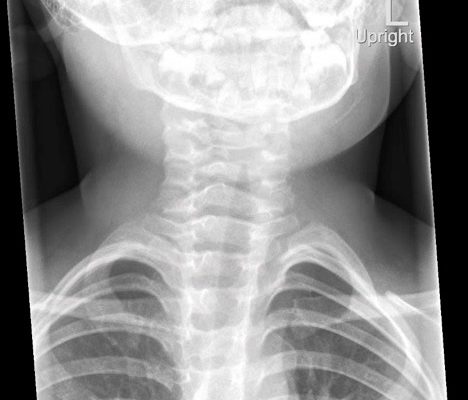
Croup
DOI: https://doi.org/10.21980/J8W05JThe anteroposterior X-ray reveals the classic steeple sign (blue outline) indicative of subglottic edema leading to tracheal narrowing, consistent with croup. The lateral x-ray shows narrowing of the subglottic region (green outline and arrows).
Various Degrees of Thermal Burns
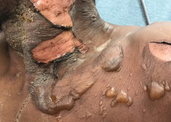
DOI: https://doi.org/10.21980/J8R91WOn exam,there is a large swath of skin with evidence of thermal injury involving the neck, shoulder, chest, and face, including damage to the ear, external nostril, and lips. Burns exhibit varying degrees of severity and total approximately 4.5% of the body surface area. Several areas are charred and insensate to pinprick. The left earlobe is partially burned off. Patient's airway is patent with no evidence of thermal injury or obstruction to the oropharynx or nasal vestibule.

