
Visual EM
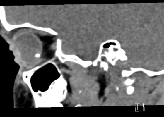

Open Globe with Intraocular Foreign Body
DOI: https://doi.org/10.21980/J8S348On physical exam, his extraocular movements were intact. The right anterior chamber appeared cloudy, particularly nasal to the pupil. The conjunctiva of the right eye was injected. The right pupil was 3 mm and sluggishly reactive and appeared slightly irregular (see yellow arrow). Of note, the right eye also had a 1 mm hypopyon, indicating inflammation of the anterior chamber, which was visible on slit lamp examination (not pictured). There was no fluorescein uptake or Seidel sign. His visual acuity was 20/60 OD (right eye) and 20/20 OS (left eye).
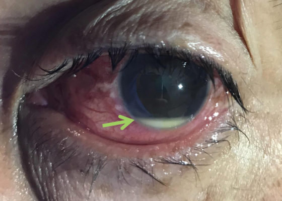
Hypopyon
DOI: https://doi.org/10.21980/J8N92BPhysical examination of the left eye revealed a hypopyon (green arrow) – which is a layered white to yellow sediment in front of the inferior aspect of the iris associated with scleral injection and chemosis. Extraocular movements were intact bilaterally and pain did not worsen with extraocular movement. The pupil was poorly reactive to direct light and only hand movement could be perceived. The intraocular pressure was 14 mmHg. Slit lamp exam demonstrated a dense cataract. Bedside ocular ultrasound demonstrated vitreous opacities concerning for possible intraocular foreign bodies.
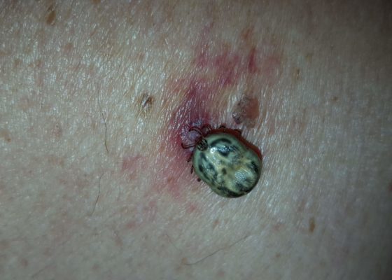
Tick Removal
DOI: https://doi.org/10.21980/J8HK9HOn physical exam, an engorged tick was found attached to the patient’s left upper back. The underlying skin was nontender but mildly erythematous, without central clearing. The tick was gently removed with blunt angle forceps and sent for further analysis, which later revealed the specimen to be an American dog tick (Dermacentor variabilis).
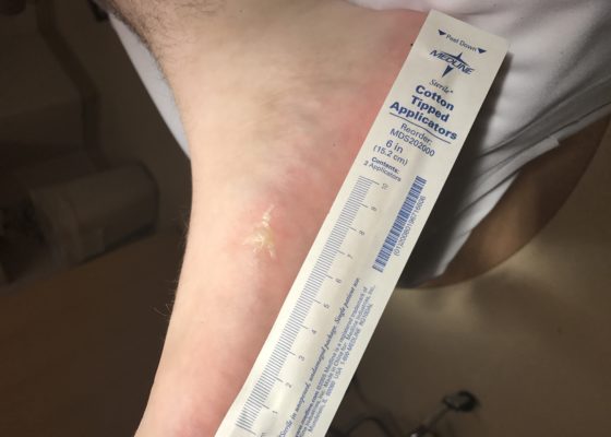
Lightning Ground Current Injury: A Subtle Shocker
DOI: https://doi.org/10.21980/J8KD1CThe first photograph demonstrates a dendritic blister (Lichtenburg figure) on the medial aspect of his right foot where the ground current injury entered the patient's foot. Although no data exists regarding the sensitivity or specificity of Lichtenberg figures as skin findings, they are considered pathognomonic for lightning injuries and are not produced by alternating current or industrial electrical injuries. The second photograph demonstrates a 4 x 3 cm area of petechiae where the ground current injury exited the patient.
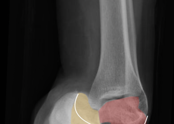
Talonavicular Dislocation
DOI: https://doi.org/10.21980/J8PG91The X-rays were significant for a subtalar dislocation. The calcaneus (red) is laterally displaced with respect to the talar head (orange), and the white lines indicate the normal articular surface. Additionally, there was a talonavicular dislocation, as seen in the fourth image: the talus (green) and navicular bone (purple) overlapping suggests a dislocation. In a normally aligned foot, the boundaries of the two bones create a point of articulation.
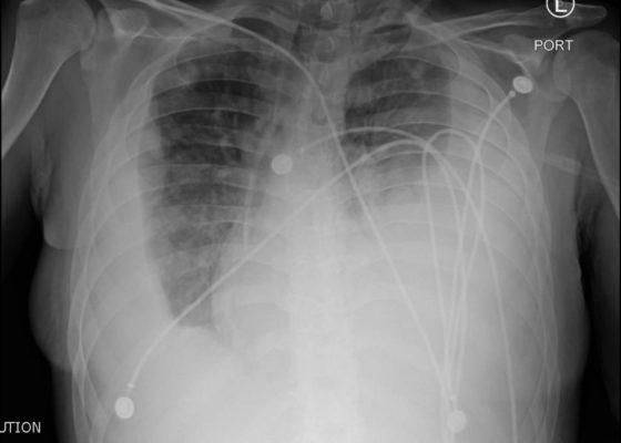
Endocarditis
DOI: https://doi.org/10.21980/J8JP73Upright frontal radiograph of the chest demonstrated large pleural effusion on the left and moderate pleural effusion on the right as shown by the visible menisci on both sides (red arrows) with diffuse bilateral nodular densities (yellow dotted lines), consistent with septic pulmonary emboli. Computed tomography (CT) of the chest demonstrated multiple scattered lung nodules bilaterally containing internal foci of air cavitation (green dotted lines).
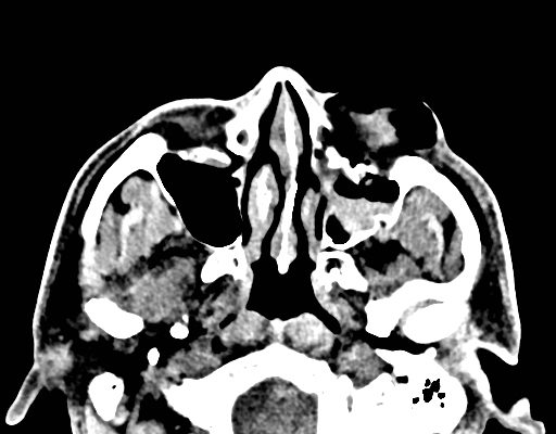
Facial Fracture Induced Periorbital Emphysema
DOI: https://doi.org/10.21980/J8F05HPhysical exam showed marked left palpebral subcutaneous crepitus, as well as bulbar and palpebral conjunctival bulging. Visual acuity was normal with intact extraocular movements, and normal pupillary exam. Computed tomography (CT) imaging of the face was obtained and revealed multiple displaced fractures involving the left orbital floor and zygomatic arch associated with moderate periorbital and postseptal extraconal gas, resulting in orbital proptosis.

Fournier Gangrene
DOI: https://doi.org/10.21980/J89626The computed tomography (CT) of the abdomen and pelvis revealed significant subcutaneous gas tracking along the perineum and right gluteal region (orange outline) into the scrotum with associated scrotal edema (yellow arrow) and subcutaneous inflammatory fat stranding of 0.92 cm (red arrow) consistent with Fournier’s gangrene. There is early fluid loculation along the right medial gluteal cleft of 5.85 cm (green arrow) without a sizeable drainable abscess seen.