• Registration
  • Login
JETem
  • Home
  • About
    • Aim and Scope
    • Our Team
    • Editorial Board
    • FAQ
  • Issues
    • Current Issue
    • Ahead of Print
    • Past Issues
  • Visual EM
    • Latest Visual EM
    • Search Visual EM
    • Thumbnail Library
  • For Authors
    • Instructions for Authors
    • Submit to JETem
    • Photo Consent
    • Policies
      • Peer Review Policy
      • Copyright Policy
      • Editorial Policy, Ethics and Responsibilities
      • Conflicts of Interest & Informed Consent
      • Open Access Policy
  • For Reviewers
    • Instructions for JETem Reviewers
    • Interested in Being a JETem Reviewer?
  • Topic
    • Abdominal / Gastroenterology
    • Administration
    • Board Review
    • Cardiology / Vascular
    • Clinical Informatics, Telehealth and Technology
    • Dermatology
    • EMS
    • Endocrine
    • ENT
    • Faculty Development
    • Genitourinary
    • Geriatrics
    • Hematology / Oncology
    • Infectious Disease
    • Miscellaneous
    • Neurology
    • Ob / Gyn
    • Ophthalmology
    • Orthopedics
    • Pediatrics
    • Pharmacology
    • Procedures
    • Psychiatry
    • Renal / Electrolytes
    • Respiratory
    • Social Determinants of Health
    • Toxicology
    • Trauma
    • Ultrasound
    • Urology
    • Wellness
    • Wilderness
  • Modality
    • Curricula
    • Innovations
    • Lectures
    • Oral Boards
      • Structured Interview
      • Communication Case
    • Podcasts
    • Simulation
    • Small Group Learning
    • Team Based Learning
    • Visual EM
  • Contact Us

Empyema

Jonathan Patane, MD* and Daniel Ritter, BS^

*University of California Irvine, Department of Emergency Medicine, Orange, CA
^The Ohio State University College of Medicine, Columbus, OH

Correspondence should be addressed to Jonathan Patane, MD at jpatane@uci.edu

DOI: https://doi.org/10.21980/J86P9RIssue 3:1
RespiratoryInfectious DiseaseVisual EM
No ratings yet.
Creative Commons images

History of present illness:

A 51-year-old male with congestive heart failure, previous myocardial infarctions, and a questionable history of chronic obstructive pulmonary disease presented to the emergency department with a chief complaint of shortness of breath. His shortness of breath developed the morning of presentation, and was associated with midsternal chest pain radiating to his right shoulder. Vital signs were significant for a temperature of 36.5ºC, heart rate of 115/minute, respiratory rate of 26/minute, and oxygen saturation of 92% on 15-liter facemask. His exam demonstrated decreased breath sounds on the right side without wheezing.  An immediate chest X-ray was performed.

Significant findings:

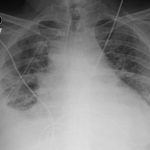
The chest X-ray shows a large fluid collection in the right lung demonstrated by the opacification that blunts the costophrenic angle on the right side. There is also a meniscus present, which is generally indicative of fluid.  Chest computed tomography (CT) demonstrated an infiltrate with a mixture of densities within the same collection, consistent with a loculated effusion and concerning for an empyema.

Discussion:

The patient’s labs were significant for an elevated white blood cell count of 20,500/mm3.  He was started on broad-spectrum antibiotics and diagnosed with an empyema. He was admitted to the ICU where he underwent thoracentesis and eventual chest tube placement.

Empyema is defined as a pleural effusion which is grossly purulent or contains bacteria visualized on gram stain.1,2  Empyemas are most commonly associated with concurrent pneumonia or invasive procedures such as thoracotomy.2  Untreated cases often develop loculations and scarring of the pleura.1 Patients with empyema typically present with fever, shortness of breath, productive cough, and/or chest pain.2  Imaging is key in diagnosis, with lateral decubitus radiographs being able to detect as little as 50 mL of fluid compared to 200 mL of fluid needed in an anteroposterior chest radiograph. 2 Ultrasound can also be used to identify loculations.  Chest CT can be used to optimally evaluate an empyema.2 Free-flowing fluid warrants sampling by thoracentesis.1,2

Empiric antibiosis should cover pseudomonas and anaerobes, with methicillin-resistant Staphylococcus aureus coverage if suspected.2 All empyemas require invasive procedures for treatment, the least invasive being chest tube placement until drainage is less than 50 mL/day.2 Unresolved empyema after chest tube placement may require video-assisted thoracoscopic surgery (VATS) or thoracotomy with decortication.1,2

Topics:

Empyema, pleural effusion, shortness of breath, chest tube, thoracentesis.

References:

  1. Light RW. Parapneumonic Effusions and empyema. Proc Am Thorac Soc. 2006;3(1):75-80. doi: 10.1513/pats.200510-113JH
  2. Desai H, Agrawal A. Pulmonary emergencies: pneumonia, acute respiratory distress syndrome, lung abscess, and empyema. Med Clin North Am. 2012;96(6):1127-1148. doi: 10.1016/j.mcna.2012.08.007
Icon

Empyema - Case Report

1 file(s) 662 KB
Download
Icon

Empyema - Images

1 file(s) 171 KB
Download
CTIssue 3:1X-Ray

Reviews:

No ratings yet.

Please rate this


Pediatric Toxic Shock Syndrome

08 Jan, 18
Creative Commons images

Acute Pyelonephritis with Perinephric Stranding...

08 Jan, 18

JETem is an online, open access, peer-reviewed, journal-repository for EM educators. We are PMC Indexed.

Most Viewed

  • The Silent Saboteur: Teaching the Clinical Implications of Occult Hypoxemia & Social Determinants of Health via a Pulmonary Embolism Case
  • Diabetic Ketoacidosis and Necrotizing Soft Tissue Infection
  • My Broken Heart
  • Stabilization of Cardiogenic Shock for Critical Care Transport, a Simulation
  • Innovative Ultrasound-Guided Erector Spinae Plane Nerve Block Model for Training Emergency Medicine Physicians

Visit Our Collaborators

Creative Commons Licence
This work is licensed under a Creative Commons Attribution 4.0 International License.

About

Education

Learners should benefit from active learning. JETem accepts submissions of team-based learning, small group learning, simulation, podcasts, lectures, innovations, curricula, question sets, and visualEM.

Scholarship

We believe educators should advance through the scholarship of their educational work. JETem gives educators the opportunity to publish scholarly academic work so that it may be widely distributed, thereby increasing the significance of their results.

Links

  • Home
  • Aim and Scope
  • Current Issue
  • For Reviewers
  • Instructions for Authors
  • Contact Us

Newsletter

Sign up to receive updates from JETem regarding newly published issues and findings.

Copyright Creative Commons Attribution 4.0 International