Posts by JETem
Little Patients, Big Tasks – A Pediatric Emergency Medicine Escape Room
DOI: https://doi.org/10.21980/J89W70By the end of this small group exercise, learners will be able to: 1) demonstrate appropriate dosing of pediatric code and resuscitation medications; 2) recognize normal pediatric vital signs by age; 3) demonstrate appropriate use of formulas to calculate pediatric equipment sizes and insertion depths; 4) recognize classic pediatric murmurs; 5) appropriately diagnose congenital cardiac conditions; 6) recognize abnormal pediatric electrocardiograms (ECGs); 7) identify life-threatening pediatric conditions; 8) demonstrate intraosseous line (IO) insertion on a pediatric model; and 9) demonstrate appropriate use of the Neonatal Resuscitation Protocol (NRP®) algorithms.
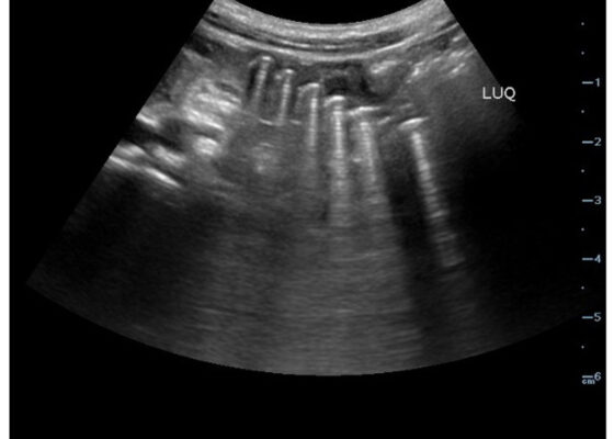
Point-Of-Care Ultrasound Use for Detection of Multiple Metallic Foreign Body Ingestion in the Pediatric Emergency Department: A Case Report
DOI: https://doi.org/10.21980/J83D2DBedside POCUS was performed on the patient’s abdomen using the curvilinear probe. The left upper quadrant POCUS image demonstrates multiple hyperechoic spherical objects with shadowing and reverberation artifacts concerning multiple foreign body ingestions. Though the patient and mother initially denied knowledge of foreign body ingestion, on repeated questioning after POCUS findings, the patient admitted to his mother that he ate the spherical magnets he received for his birthday about one week ago. The patient swallowed these over the course of two days. The presence of multiple radiopaque foreign bodies was confirmed with an abdominal X-ray.
Sonographic Retrobulbar Spot Sign in Diagnosis of Central Retinal Artery Occlusion: A Case Report
DOI: https://doi.org/10.21980/J8735PThe bedside ocular ultrasound (B-scan) was significant for small, hyperechoic signal (white arrow) in the distal aspect of the optic nerve, concerning for embolus in the central retinal artery. Subsequent direct fundoscopic exam was significant for a pale macula with cherry red spot (black arrow), consistent with central retinal artery occlusion (CRAO).
Everyday Water-Related Emergencies: A Didactic Course Expanding Wilderness Medicine Education
DOI: https://doi.org/10.5072/FK2HX1GX76By the end of the session, the learner will be able to: 1) describe the pathophysiology of drowning and shallow water drowning, 2) prevent water emergencies by listing water preparations and precautions to take prior to engaging in activities in and around water, 3) recognize a person at risk of drowning and determine the next best course of action, 4) demonstrate three different methods for in-water c-spine stabilization in the case of a possible cervical injury, 5) evaluate and treat a patient after submersion injury, 6) appropriately place a tourniquet for hemorrhage control, and 7) apply a splint to immobilize skeletal injury.
Alcohol Withdrawal with Delirium Tremens
DOI: https://doi.org/10.21980/J8S35NBy the end of the session, learner will be able to 1) discuss the causes of altered mental status, 2) utilize CIWA scoring system to quantify AW severity, 3) formulate appropriate treatment plan for AW by treating with benzodiazepine and escalating treatment appropriately, 4) treat electrolyte abnormalities by giving appropriate medications for hypokalemia and hypomagnesemia, and 5) discuss clinical progression and timing to AW.
Headache Over Heels: CT Negative Subarachnoid Hemorrhage
DOI: https://doi.org/10.21980/J8ND2CBy the end of this case, the participant will be able to: 1) construct a broad differential diagnosis for a patient presenting with syncope, 2) name the history and physical exam findings consistent with SAH, 3) identify SAH on computer tomography (CT) imaging, 4) identify the need for lumbar puncture (LP) to diagnose SAH when CT head is non-diagnostic > 6 hours after symptom onset, 5) correctly interpret cerebral fluid studies (CSF) to aid in the diagnosis of SAH, and 6) specify blood pressure goals in SAH and suggest appropriate medication management.
A Homemade, Cost-Effective, Realistic Pelvic Exam Model
DOI: https://doi.org/10.21980/J8HM0FAfter utilizing this pelvic examination model, the learner will be able to: 1) demonstrate ability to perform a pelvic examination comfortably and safely, 2) demonstrate ability to obtain a cervical swab on female patients, and 3) show proficient understanding of female anatomy.


