• Registration
  • Login
JETem
  • Home
  • About
    • Aim and Scope
    • Our Team
    • Editorial Board
    • FAQ
  • Issues
    • Current Issue
    • Ahead of Print
    • Past Issues
  • Visual EM
    • Latest Visual EM
    • Search Visual EM
    • Thumbnail Library
  • For Authors
    • Instructions for Authors
    • Submit to JETem
    • Photo Consent
    • Policies
      • Peer Review Policy
      • Copyright Policy
      • Editorial Policy, Ethics and Responsibilities
      • Conflicts of Interest & Informed Consent
      • Open Access Policy
  • For Reviewers
    • Instructions for JETem Reviewers
    • Interested in Being a JETem Reviewer?
  • Topic
    • Abdominal / Gastroenterology
    • Administration
    • Board Review
    • Cardiology / Vascular
    • Clinical Informatics, Telehealth and Technology
    • Dermatology
    • EMS
    • Endocrine
    • ENT
    • Faculty Development
    • Genitourinary
    • Geriatrics
    • Hematology / Oncology
    • Infectious Disease
    • Miscellaneous
    • Neurology
    • Ob / Gyn
    • Ophthalmology
    • Orthopedics
    • Pediatrics
    • Pharmacology
    • Procedures
    • Psychiatry
    • Renal / Electrolytes
    • Respiratory
    • Social Determinants of Health
    • Toxicology
    • Trauma
    • Ultrasound
    • Urology
    • Wellness
    • Wilderness
  • Modality
    • Curricula
    • Innovations
    • Lectures
    • Oral Boards
      • Structured Interview
      • Communication Case
    • Podcasts
    • Simulation
    • Small Group Learning
    • Team Based Learning
    • Visual EM
  • Contact Us

Issue 1:1

Creative Commons images

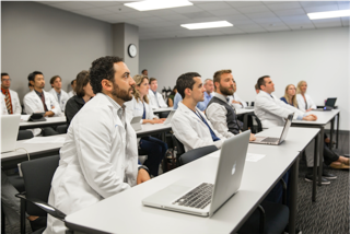
A Faculty Development Session or Resident as Teacher Session for Didactic and Clinical Teaching Techniques; Part 1 of 2: Engaging Learners with Effective Didactic Teaching

Megan Boysen-Osborn, MD*, Shannon Toohey, MD*, Michael Gisondi, MD‡, and Margaret Wolff , MD^

DOI: https://doi.org/10.21980/J8RP4TIssue 1:1 5/5 (1)
By the end of this workshop, the learner will: 1) describe eight teaching techniques that encourage active learning during didactic sessions; 2) plan a didactic session using at least one of eight new teaching techniques for didactic instruction
Faculty DevelopmentLecturesTeam Based Learning (TBL)
Creative Commons images

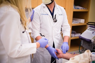
A Faculty Development Session or Resident as Teacher Session for Clinical and Clinical Teaching Techniques; Part 2 of 2: Engaging Learners with Effective Clinical Teaching

Megan Boysen-Osborn, MD*, Margaret Wolff, MD‡, Michael Gisondi, MD^

DOI: https://doi.org/10.21980/J8MW2WIssue 1:1 No ratings yet.
By the end of this workshop, the learner will: 1) describe and implement nine new clinical teaching techniques; 2) implement clinical teaching techniques specific to junior and senior resident learners.
Faculty DevelopmentLecturesTeam Based Learning (TBL)

Carbon Monoxide Poisoning

Alisa Wray, MD*

DOI: https://doi.org/10.21980/J8KH59CIssue 1:1 No ratings yet.
By completing this oral board case learners will 1) evaluate a patient with altered mental status and discuss the differential diagnosis of a patient with altered mental status and weakness; 2) recognize the signs and symptoms of carbon monoxide poisoning; 3) manage treatment of a patient with carbon monoxide poisoning
ToxicologyBoard ReviewOral Boards

Emergencies in Hemodialysis Patients

Shannon Toohey, MD*

DOI: https://doi.org/10.21980/J81591Issue 1:1 No ratings yet.
By the end of this session, the learner will: 1) describe primary dialysis complications; 2) construct a full differential for a dialysis patient presenting with complications; 3) formulate an appropriate treatment and resuscitation in an acutely ill dialysis patient; 4) plan appropriate disposition and utilization of consultants for dialysis complications.
Renal/ElectrolytesTeam Based Learning (TBL)

Approach to Acute Headache: A Flipped Classroom Module for Emergency Medicine Trainees

Jeff Riddell, MD*, Stacy Sawtelle, MD^, Paul Jhun, MD‡, James Comes, MD^, Ramin Tabatabai, MD~, Daniel Joseph, MD~, Jan Shoenberger, MD~, Esther Chen, MD‡, Christopher Fee, MD‡, and Stuart Swadron, MD~

DOI: https://doi.org/10.21980/J8WC73Issue 1:1 No ratings yet.
At the end of this module, the learner will be able to: 1) list the diagnoses critical to the emergency physician that may present with headache; 2) identify key historical and examination findings that help differentiate primary (benign) from secondary (serious) causes of headache; 3) discuss the indications for diagnostic imaging, lumbar puncture and laboratory testing in patients with headache; 4) recognize life-threatening diagnoses on CT imaging and CSF examination; 5) describe treatment strategies to relieve headache symptoms.
NeurologySmall Group Learning

Mesenteric Ischemia

Shannon Toohey, MD*

DOI: https://doi.org/10.21980/J8CC7FIssue 1:1 No ratings yet.
At the end of this simulation session, the learner will: 1) Recognize signs and symptoms of mesenteric ischemia; 2) order appropriately imaging and labs in the workup of an elderly patient with abdominal pain; 3) manage a patient with mesenteric ischemia, a rare, but serious cause of abdominal pain in the elderly; 4) discuss anchoring bias, specifically related to patients referred to the ED with an established diagnosis by outside specialists.
Abdominal/GastroenterologySimulation
Creative Commons images

Atrial Myxoma

Alisa Wray, MD*

DOI: https://doi.org/10.21980/J87P45Issue 1:1 No ratings yet.
Bedside ultrasound revealed the presence of a left atrial mass that appeared to be tethered to the mitral valve. The mass was best viewed on ultrasound in the apical four-chamber window with the phased array probe placed over the patients’ point of maximal impact (PMI), with the patient in left lateral decubitus position.
UltrasoundCardiology/VascularVisual EM
Creative Commons images

Hampton’s Hump in Pulmonary Embolism

Jonathan Patane, MD* and Megan Boysen Osborn*

DOI: https://doi.org/10.21980/J83W27Issue 1:1 No ratings yet.
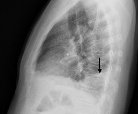
In the lateral view chest x-ray, there is a Hampton’s Hump, a pleural based, wedge-shaped opacity at the base of the right lung, representing lung infarction (black arrow). These findings correlate with the sagittal view on CT angiography of the chest. The CT chest also shows a filling defect in the distal posterior basal segmental pulmonary artery (white arrow), as demonstrated by the absence of contrast enhancement in the distal portion of the vessel. This is associated with an opacification of the lung parenchyma distal to the occlusion (red arrow), representing lung infarction.
RespiratoryVisual EM
Creative Commons images

Acute Pericarditis: Electrocardiogram

Jason Mefford, MD* and Shannon L Toohey, MD*

DOI: https://doi.org/10.21980/J8059QIssue 1:1 No ratings yet.
The ECG shows diffuse ST- elevation. The patient also has mild PR-depression, most notably in the inferior and lateral leads. The patient also has minimal PR elevation in lead aVR. The patient was diagnosed with acute pericarditis (ECG stage 1).
Cardiology/VascularVisual EM
Creative Commons images

Thompson Test in Achilles Tendon Rupture

Spencer Albertson* and Megan Boysen Osborn, MD, MHPE*

DOI: https://doi.org/10.21980/J8VC7SIssue 1:1 No ratings yet.
The left Achilles tendon had a defect on palpation, while the right Achilles tendon was intact. When squeezing the right (unaffected) calf, the ankle spontaneously plantar flexed, indicating a negative (normal) Thompson test. Upon squeeze of the left (affected) calf, the ankle did not plantar flex, signifying a positive (abnormal) Thompson test. The diagnosis of left Achilles tendon rupture was confirmed intraoperatively one week later.
OrthopedicsTraumaVisual EM

JETem is an online, open access, peer-reviewed, journal-repository for EM educators. We are PMC Indexed.

Most Viewed

  • The Silent Saboteur: Teaching the Clinical Implications of Occult Hypoxemia & Social Determinants of Health via a Pulmonary Embolism Case
  • Diabetic Ketoacidosis and Necrotizing Soft Tissue Infection
  • My Broken Heart
  • Stabilization of Cardiogenic Shock for Critical Care Transport, a Simulation
  • Innovative Ultrasound-Guided Erector Spinae Plane Nerve Block Model for Training Emergency Medicine Physicians

Visit Our Collaborators

Creative Commons Licence
This work is licensed under a Creative Commons Attribution 4.0 International License.

About

Education

Learners should benefit from active learning. JETem accepts submissions of team-based learning, small group learning, simulation, podcasts, lectures, innovations, curricula, question sets, and visualEM.

Scholarship

We believe educators should advance through the scholarship of their educational work. JETem gives educators the opportunity to publish scholarly academic work so that it may be widely distributed, thereby increasing the significance of their results.

Links

  • Home
  • Aim and Scope
  • Current Issue
  • For Reviewers
  • Instructions for Authors
  • Contact Us

Newsletter

Sign up to receive updates from JETem regarding newly published issues and findings.

Copyright Creative Commons Attribution 4.0 International