• Registration
  • Login
JETem
  • Home
  • About
    • Aim and Scope
    • Our Team
    • Editorial Board
    • FAQ
  • Issues
    • Current Issue
    • Ahead of Print
    • Past Issues
  • Visual EM
    • Latest Visual EM
    • Search Visual EM
    • Thumbnail Library
  • For Authors
    • Instructions for Authors
    • Submit to JETem
    • Photo Consent
    • Policies
      • Peer Review Policy
      • Copyright Policy
      • Editorial Policy, Ethics and Responsibilities
      • Conflicts of Interest & Informed Consent
      • Open Access Policy
  • For Reviewers
    • Instructions for JETem Reviewers
    • Interested in Being a JETem Reviewer?
  • Topic
    • Abdominal / Gastroenterology
    • Administration
    • Board Review
    • Cardiology / Vascular
    • Clinical Informatics, Telehealth and Technology
    • Dermatology
    • EMS
    • Endocrine
    • ENT
    • Faculty Development
    • Genitourinary
    • Geriatrics
    • Hematology / Oncology
    • Infectious Disease
    • Miscellaneous
    • Neurology
    • Ob / Gyn
    • Ophthalmology
    • Orthopedics
    • Pediatrics
    • Procedures
    • Psychiatry
    • Renal / Electrolytes
    • Respiratory
    • Toxicology
    • Trauma
    • Ultrasound
    • Wellness
    • Wilderness
  • Modality
    • Curricula
    • Innovations
    • Lectures
    • Oral Boards
    • Podcasts
    • Simulation
    • Small Group Learning
    • Team Based Learning
    • Visual EM
  • Contact Us

Hampton’s Hump in Pulmonary Embolism

Jonathan Patane, MD* and Megan Boysen Osborn*

*University of California, Irvine, Department of Emergency Medicine, Orange, CA

Correspondence should be addressed to Jonathan Patane, MD at jpatane@uci.edu

DOI: https://doi.org/10.21980/J83W27Issue 1:1
RespiratoryVisual EM
No ratings yet.


HPI:

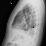
A 51-year old male presented to the emergency department with right chest and flank pain, with intermittent cough productive of blood-tinged sputum. The patient denied shortness of breath. The patient was afebrile, had a heart rate of 104/min, respiratory rate of 18/min, and oxygen saturation of 98% on room air.  Cardiopulmonary exam was unremarkable. Chest radiograph (shown) was concerning for Hampton’s hump. Subsequently, the diagnosis of pulmonary embolism was confirmed on CT angiography of the chest (shown).

Significant Findings:

In the lateral view chest x-ray, there is a Hampton’s Hump, a pleural based, wedge-shaped opacity at the base of the right lung, representing lung infarction (black arrow). These findings correlate with the sagittal view on CT angiography of the chest. The CT chest also shows a filling defect in the distal posterior basal segmental pulmonary artery (white arrow), as demonstrated by the absence of contrast enhancement in the distal portion of the vessel. This is associated with an opacification of the lung parenchyma distal to the occlusion (red arrow), representing lung infarction.

Discussion:

The diagnosis of pulmonary embolism is typically made by CT pulmonary angiography, but other diagnostic modalities include: ventilation-perfusion scan, d-dimer, ultrasound of the lower extremities, echocardiogram, electrocardiogram, chest radiograph, and clinical decision rules. Hampton’s hump (figure 1), a wedge-shaped consolidation with the base of the triangle against the pleura, was first described on the chest x-ray in the 1940s and is one finding that may be present on chest radiography.1-3

The presence of Hampton’s hump possesses a high specificity (82%), but has a low sensitivity (22%) for the diagnosis of pulmonary embolism.3,4  In patients with Hampton’s hump, CTA will confirm the diagnosis, showing a three-dimensional infarction distal to the embolus.

Topics:

Pulmonary embolism (PE), chest x-ray (CXR), CT angiography, respiratory, pulmonary, lung, emergency medicine, Hampton’s hump, pulmonary infarction.

References:

  1. Hampton AO, Castleman B. Correlation of postmortem chest teleroentgenograms with autopsy findings with special reference to pulmonary embolism and infarction. AJR Am J Roentgenol. 1940;43:305-326.
  2. Marshall GB, Farnquist BA, MacGregor JH, Burrowes PW. Signs in thoracic imaging. J Thorac Imaging. 2006;21(1):76-90. doi: 10.1097/01.rti.0000189192.70442.7a
  3. Worsley DF, Alavi A, Aronchick JM, Chen JT, Greenspan RH, Ravin CE. Chest radiographic findings in patients with acute pulmonary embolism: observations from the PIOPED Study. Radiology. 1993;189(1):133–136. doi: 10.1148/radiology.189.1.8372182
  4. Daehee H, Kyung SL, Tomas F, et al. Thrombotic and nonthrombotic pulmonary arterial embolism: spectrum of imaging findings. RadioGraphics. 2003;23(6):1521-1539. doi: 10.1148/rg.1103035043
Icon

Hampton’s Hump in Pulmonary Embolism - Case Report

1 file(s) 697 KB
Download
Icon

Hampton’s Hump in Pulmonary Embolism - Images

1 file(s) 166 KB
Download
Issue 1:1X-Ray

Reviews:

No ratings yet.

Please rate this


Acute Pericarditis: Electrocardiogram

24 Jun, 16

Atrial Myxoma

24 Jun, 16
JETem is an online, open access, peer-reviewed journal-repository for EM educators

Most Viewed

  • Telemedicine Consult for Shortness of Breath Due to Sympathetic Crashing Acute Pulmonary Edema
  • Anticholinergic Toxicity in the Emergency Department
  • The Suicidal Patient in the Emergency Department Team-Based Learning Activity
  • Child Maltreatment Education: Utilizing an Escape Room Activity to Engage Learners on a Sensitive Topic
  • Acute Chest Syndrome

Visit Our Collaborators

About

Education

Learners should benefit from active learning. JETem accepts submissions of team-based learning, small group learning, simulation, podcasts, lectures, innovations, curricula, question sets, and visualEM.

Scholarship

We believe educators should advance through the scholarship of their educational work. JETem gives educators the opportunity to publish scholarly academic work so that it may be widely distributed, thereby increasing the significance of their results.

Links

  • Home
  • Aim and Scope
  • Current Issue
  • For Reviewers
  • Instructions for Authors
  • Contact Us

Newsletter

Sign up to receive updates from JETem regarding newly published issues and findings.

Copyright © 2016 JETem. All rights reserved.