Abdominal/Gastroenterology
Incidental Hiatal Hernia on Chest X-ray
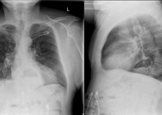
DOI: https://doi.org/10.21980/J8KP8SThe two-view chest X-ray shows mild opacification of the bilateral lower lobes concerning for pneumonia (red arrows). Incidental retrocardiac opacity with air-fluid level consistent with large hiatal hernia is also observed (green arrow).
Button Battery in Esophagus
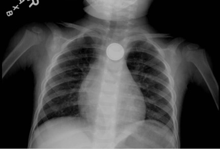
DOI: https://doi.org/10.21980/J8FW6VChest radiograph showed the presence of a round radiopaque foreign body in the mid-chest. It was suspected to be in the esophagus rather than in the trachea due to the en-face positioning of the foreign body. The foreign body demonstrated two concentric ring circles concerning for a “double ring” or “halo" sign, which was suggestive of the presence of a button battery rather than a coin.
Achalasia: An Uncommon Presentation with Classic Imaging
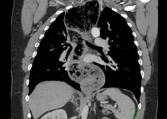
DOI: https://doi.org/10.21980/J86D2BThe chest X-ray demonstrated a markedly widened mediastinum (red brackets), raising concern for thoracic aortic aneurysm/aortic dissection, which prompted labs and contrast-enhanced computed tomography (CT) of the chest. The CT revealed a dilated proximal esophagus that narrowed distally (yellow tracing and red arrow), with particulate material, mass-effect on the trachea (purple outline), and bilateral patchy opacities suggesting aspiration. Barium esophagram showed a drastically dilated esophagus filled with contrast (yellow arrow), terminating into the classic “bird’s beak sign” (red arrow) at the lower esophageal sphincter (LES). Esophageal manometry later confirmed achalasia, proving that widened mediastina can have unexpected etiologies.
Point of Care Ultrasound Illustrating Small Bowel Obstruction
DOI: https://doi.org/10.21980/J8T637POCUS of the small bowel illustrated significantly dilated loops of bowel (white line), thickened bowel wall (white arrow) and to-and-fro peristalsis, consistent with small bowel obstruction.
Sepsis Secondary to an Abdominal Wound Infection
DOI: https://doi.org/10.21980/J8PS60At completion of this case learners should be able to: 1) Recognize and differentiate between systemic inflammatory response syndrome, sepsis, severe sepsis, and septic shock. 2) Prepare an appropriate differential diagnosis for a patient with sepsis. 3) Demonstrate appropriate fluid resuscitation and antibiotic therapy for a septic patient. 4) Demonstrate appropriate vasopressor therapy for a septic patient. 5) Understand and apply the Surviving Sepsis Guidelines.
Intussusception
DOI: https://doi.org/10.21980/J8SH0WA segment of bowel within the right abdomen that measured approximately 1.6 x 1.5 cm transaxially. It demonstrated a hypoechoic edematous outer loop of bowel (blue arrow) and hyperechoic compressed loop of bowel telescoping within (red star), this is known as the "target sign."
Procedural Sedation for the removal of a rectal foreign body
DOI: https://doi.org/10.21980/J81332Axial and coronal views on CT showed evidence of a large, tube-shaped foreign body in the rectum (see arrows) without evidence of acute gastrointestinal tract disease.
Large Ventral Hernia
DOI: https://doi.org/10.21980/J86K9QComputed tomography (CT) scan with intravenous (IV) contrast of the abdomen and pelvis demonstrated a large pannus containing a ventral hernia with abdominal contents extending below the knees (white circle), elongation of mesenteric vessels to accommodate abdominal contents outside of the abdomen (white arrow) and air fluid levels (white arrow) indicating a small bowel obstruction.
Elderly female with acute abdominal pain presenting with Superior Mesenteric Artery Thrombus
DOI: https://doi.org/10.21980/J82W52Computed tomography (CT) angiogram of the abdomen and pelvis revealed a superior mesenteric artery (SMA) thrombosis 5 cm from the origin off of the abdominal aorta. As seen in the sagittal view, there does not appear to be any contrast 5 cm past the origin of the SMA. On the axial views, you can trace the SMA until the point that there is no longer any contrast visible which indicates the start of the thrombus. The SMA does not appear to be reconstituted. There was normal flow to the celiac artery. (See annotated images).
A Case of Acute Cholecystitis
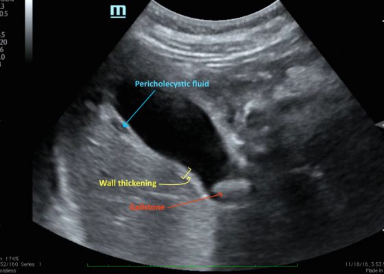
DOI: https://doi.org/10.21980/J8405QBedside point-of-care ultrasound revealed a distended gallbladder, thickened gallbladder wall, pericholecystic fluid, and a stone in the neck of the gallbladder indicative of acute cholecystitis.


