
JETem
  • Home
  • About
    • Aim and Scope
    • Our Team
    • Editorial Board
    • FAQ
  • Issues
    • Current Issue
    • Ahead of Print
    • Past Issues
  • Visual EM
    • Latest Visual EM
    • Search Visual EM
    • Thumbnail Library
  • For Authors
    • Instructions for Authors
    • Submit to JETem
    • Photo Consent
    • Policies
      • Peer Review Policy
      • Copyright Policy
      • Editorial Policy, Ethics and Responsibilities
      • Conflicts of Interest & Informed Consent
      • Open Access Policy
      • Acceptance Fee
  • For Reviewers
    • Instructions for JETem Reviewers
    • Interested in Being a JETem Reviewer?
  • Topic
    • Abdominal/Gastroenterology
    • Administration
    • Board Review
    • Cardiology/Vascular
    • Certifying Exam Cases
      • Clinical Decision-Making
      • Prioritization
      • Communication
      • Procedure & Ultrasound
    • Clinical Informatics, Telehealth and Technology
    • Dermatology
    • EMS
    • Endocrine
    • ENT
    • Faculty Development
    • Genitourinary
    • Geriatrics
    • Hematology/Oncology
    • Infectious Disease
    • Miscellaneous (stats, etc)
    • Neurology
    • Ob/Gyn
    • Ophthalmology
    • Orthopedics
    • Pediatrics
    • Pharmacology
    • Procedures
    • Psychiatry
    • Renal/Electrolytes/Nutrition
    • Respiratory
    • Social Determinants of Health
    • Toxicology
    • Trauma
    • Ultrasound
    • Urology
    • Wellness
    • Wilderness
  • Modality
    • Certifying Exam Practice Cases
      • Clinical Care Cases
      • Communication & Procedural Cases
      • Old School Oral Board Cases
    • Curricula
    • Innovations
    • Lectures
    • Podcasts
    • Simulation
    • Small Group Learning
    • Team Based Learning (TBL)
    • Visual EM
  • Contact Us

Abdominal/Gastroenterology
Creative Commons images

Achalasia: An Uncommon Presentation with Classic Imaging

Joseph Adamson, BS*, Mina Altwail, MD^ and Shanna Jones, MD^

DOI: https://doi.org/10.21980/J86D2BIssue 3:3[mrp_rating_result]
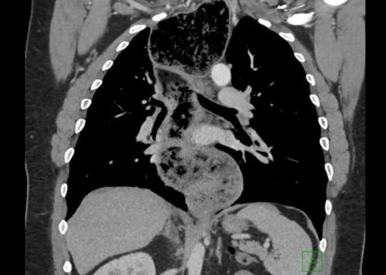
The chest X-ray demonstrated a markedly widened mediastinum (red brackets), raising concern for thoracic aortic aneurysm/aortic dissection, which prompted labs and contrast-enhanced computed tomography (CT) of the chest. The CT revealed a dilated proximal esophagus that narrowed distally (yellow tracing and red arrow), with particulate material, mass-effect on the trachea (purple outline), and bilateral patchy opacities suggesting aspiration. Barium esophagram showed a drastically dilated esophagus filled with contrast (yellow arrow), terminating into the classic “bird’s beak sign” (red arrow) at the lower esophageal sphincter (LES). Esophageal manometry later confirmed achalasia, proving that widened mediastina can have unexpected etiologies.
Abdominal/GastroenterologyVisual EM
Creative Commons images

Point of Care Ultrasound Illustrating Small Bowel Obstruction

Badriah Alruwaili, MBBS* and Shadi Lahham, MD*

DOI: https://doi.org/10.21980/J8T637Issue 3:3[mrp_rating_result]
POCUS of the small bowel illustrated significantly dilated loops of bowel (white line), thickened bowel wall (white arrow) and to-and-fro peristalsis, consistent with small bowel obstruction.
Abdominal/GastroenterologyUltrasoundVisual EM

Sepsis Secondary to an Abdominal Wound Infection

Alisa Wray, MD*

DOI: https://doi.org/10.21980/J8PS60 Issue 3:2[mrp_rating_result]
At completion of this case learners should be able to: 1) Recognize and differentiate between systemic inflammatory response syndrome, sepsis, severe sepsis, and septic shock. 2) Prepare an appropriate differential diagnosis for a patient with sepsis. 3) Demonstrate appropriate fluid resuscitation and antibiotic therapy for a septic patient. 4) Demonstrate appropriate vasopressor therapy for a septic patient. 5) Understand and apply the Surviving Sepsis Guidelines.
Infectious DiseaseAbdominal/GastroenterologySimulation
Creative Commons images

Intussusception

Julian Jakubowski, DO*

DOI: https://doi.org/10.21980/J8SH0WIssue 3:2[mrp_rating_result]
A segment of bowel within the right abdomen that measured approximately 1.6 x 1.5 cm transaxially. It demonstrated a hypoechoic edematous outer loop of bowel (blue arrow) and hyperechoic compressed loop of bowel telescoping within (red star), this is known as the "target sign."
Abdominal/GastroenterologyVisual EM
Creative Commons images

Procedural Sedation for the removal of a rectal foreign body

John Costumbrado, MD, MPH*, Valerie Lew, BS* and David Wagner, MD*

DOI: https://doi.org/10.21980/J81332Issue 3:2[mrp_rating_result]
Axial and coronal views on CT showed evidence of a large, tube-shaped foreign body in the rectum (see arrows) without evidence of acute gastrointestinal tract disease.
Abdominal/GastroenterologyProceduresVisual EM
Creative Commons images

Large Ventral Hernia

Meryl Abrams, MD*, Raymond Feuga, MD*, Nicholas Governatori, MD* and Jennifer White, MD*

DOI: https://doi.org/10.21980/J86K9QIssue 3:2[mrp_rating_result]
Computed tomography (CT) scan with intravenous (IV) contrast of the abdomen and pelvis demonstrated a large pannus containing a ventral hernia with abdominal contents extending below the knees (white circle), elongation of mesenteric vessels to accommodate abdominal contents outside of the abdomen (white arrow) and air fluid levels (white arrow) indicating a small bowel obstruction.
Abdominal/GastroenterologyVisual EM
Creative Commons images

Elderly female with acute abdominal pain presenting with Superior Mesenteric Artery Thrombus

Sassan Ghassemzadeh, MD*

DOI: https://doi.org/10.21980/J82W52 Issue 3:2[mrp_rating_result]
Computed tomography (CT) angiogram of the abdomen and pelvis revealed a superior mesenteric artery (SMA) thrombosis 5 cm from the origin off of the abdominal aorta. As seen in the sagittal view, there does not appear to be any contrast 5 cm past the origin of the SMA. On the axial views, you can trace the SMA until the point that there is no longer any contrast visible which indicates the start of the thrombus.  The SMA does not appear to be reconstituted. There was normal flow to the celiac artery. (See annotated images).
Abdominal/GastroenterologyCardiology/VascularVisual EM
Creative Commons images

A Case of Acute Cholecystitis

Chad Correa, BS* and Lindsey Spiegelman, MD^

DOI: https://doi.org/10.21980/J8405QIssue 3:1[mrp_rating_result]
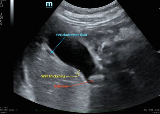
Bedside point-of-care ultrasound revealed a distended gallbladder, thickened gallbladder wall, pericholecystic fluid, and a stone in the neck of the gallbladder indicative of acute cholecystitis.
Abdominal/GastroenterologyVisual EM
«‹7›»
Page 7 of 10

JETem is an online, open access, peer-reviewed, journal-repository for EM educators. We are PMC Indexed.

Most Viewed

  • Simple Auricular Hematoma (“Cauliflower Ear”) Task Trainer
  • The Three Breath Method: Teaching Coping Mechanisms for Processing Patient Death
  • Clinical Decision Making: Acute Ischemic Stroke with Large Vessel Occlusion
  • PRIMER: Premedical Introduction to Mentored Emergency Research: Connecting Undergraduate Assistants and Emergency Medicine Residents in a Community Hospital Setting
  • Dangerous Dysrhythmias in the ED: An Interactive EKG Workshop for Emergency Medicine Clerkship Learners

Visit Our Collaborators


Creative Commons Licence
This work is licensed under a Creative Commons Attribution 4.0 International License.

About

Education

Learners should benefit from active learning. JETem accepts submissions of team-based learning, small group learning, simulation, podcasts, lectures, innovations, curricula, question sets, and visualEM.

Scholarship

We believe educators should advance through the scholarship of their educational work. JETem gives educators the opportunity to publish scholarly academic work so that it may be widely distributed, thereby increasing the significance of their results.

Links

  • Home
  • Aim and Scope
  • Current Issue
  • For Reviewers
  • Instructions for Authors
  • Contact Us

Newsletter

Sign up to receive updates from JETem regarding newly published issues and findings.

Copyright Creative Commons Attribution 4.0 International