Photograph
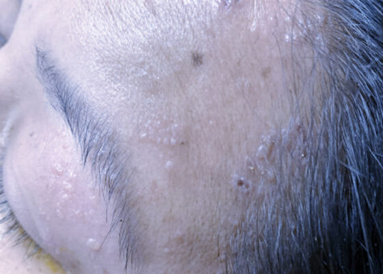
Case Report of Herpes Zoster Ophthalmicus with Concurrent Parotitis
DOI: https://doi.org/10.21980/J8R93NThe presence of soft tissue stranding about the parotid gland suggested an underlying inflammatory or infectious process of the parotid gland. Cellulitis was considered as a possible diagnosis as well, given the presence of soft tissue stranding in the dermis that is adjacent to the parotid gland. Fortunately, no enhancement was seen in local muscles, fascia, or bones to suggest a myositis, fasciitis, or osteomyelitis. By using the anatomy of the patient and understanding the changes that occur on CT when inflammation is present, the appropriate depth and location of infection can be made, allowing for appropriate treatment regimens.
A Man with Sore Throat—A Case Report
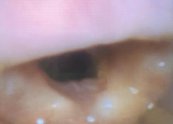
DOI: https://doi.org/10.21980/J8MH0BVideo laryngoscopy of the upper airway was performed two days after initial burn injury. The images obtained demonstrated laryngeal edema and inflammation near the epiglottis. The dot identifies the epiglottis and the asterix identifies the area of moderate thermal burns. Imaging also demonstrated adequate patency of airway and ruled out the need for intubation at that time.
The Continued Rise of Syphilis: A Case Report to Aid in Identification of the Great Imitator
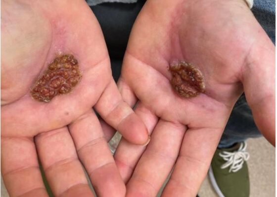
DOI: https://doi.org/10.21980/J8R93NImages taken of the bilateral palmar skin lesions at our institution showed multi-centimeter, well-demarcated, friable, verrucous, crusted plaques with overlying fine yellow crust. Lesions such as these are suspicious for syphilitic gummas seen with cutaneous tertiary syphilis.
Case Report of a Tongue-Type Calcaneal Fracture
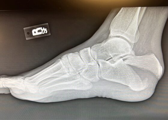
DOI: https://doi.org/10.21980/J8NH11Examination of the right ankle demonstrated a large deformity of the superior talus with bruising and blanching of the overlying skin in the area of the Achilles tendon (see images 2,3). The remaining bones of the foot were not tender to palpation and the foot was neurovascularly intact throughout with only mild numbness in the area of the tented skin. Completing the trauma exam, the patient had no signs of head injury and no midline spinal tenderness to palpation. Inspection of the remaining long bones and joints showed no other injuries. There were mild skin scrapes on the right flank from the fall. X-rays of the right foot and ankle showed a longitudinal fracture of the calcaneal tuberosity from the articular surface to the posterior surface (see red outline) with extension into the subtalar joint (blue lines) and roughly 1.8 cm displacement between the fracture segments (yellow double arrow). These findings represented a tongue-type calcaneal bone fracture.
Mushroom for Improvement Case Report: The Importance of Involving Mycologists
DOI: https://doi.org/10.21980/J8ZW7WThe mushroom displayed here is large and lacks any gills. Small puffball mushrooms can resemble young immature button top Amanita type mushrooms. Opening the Amanita mushroom should reveal apparent gills and quickly differentiate the two- -the puffball mushroom should have a white interior without gills.
Evaluation of ACE-inhibitor Induced Laryngeal Edema Using Fiberoptic Scope: A Case Report
DOI: https://doi.org/10.21980/J83P9TPhysical exam was initially significant for swelling isolated to the right sided cheek and upper lip. There was no edema to lower lip, uvular swelling, or swelling to the submandibular space. She was speaking full sentences and did not endorse any voice changes. Initial vital signs were as follows: BP 125/77, HR 74, RR 16, and oxygen saturation of 100% on room air. Approximately 40 minutes later, after 125 mg solumedrol intravenous (IV) and 50mg diphenhydramine by mouth, swelling had spread to the entire upper lip and the patient reported spreading to her jaw (Photo 1). Although no jaw or submandibular edema was appreciated on physical exam, a flexible fiberoptic laryngoscope was used to evaluate the patient’s airways given worsening symptoms. Viscous lidocaine was applied intranasally five minutes prior to the procedure. The patient was positioned in a seated position on the stretcher. A flexible fiberoptic laryngoscope was then inserted through the nares and advanced slowly. Laryngoscopy showed diffuse edema of the epiglottis, arytenoids, and ventricular folds (see photos 2-4). Vital signs and respiratory status remained stable both during and after the procedure.
A Case Report of Fournier’s Gangrene
DOI: https://doi.org/10.21980/J8Z356Physical exam revealed a comfortable-appearing male patient with tachycardia and a regular cardiac rhythm. The genitourinary exam indicated significant erythema and fluctuance of the bilateral lower buttocks with extension to the perineum. Black eschar and ecchymosis were also noted at the perineum. There was significant tenderness to palpation that extended beyond the borders of erythema. There was no palpable crepitus on initial examination. Physical exam was otherwise unremarkable.
A Case Report of the Rapid Evaluation of a High-Pressure Injection Injury of a Finger Leading to Positive Outcomes
DOI: https://doi.org/10.21980/J8TD2XOn exam the patient was noted to have a punctate wound to the ulnar aspect of his right index finger, just proximal to the distal interphalangeal joint. The finger appeared pale and taut, with absent capillary refill. The patient displayed diminished range of motion with both extension and flexion of the joints of the finger. Sensation was absent and no doppler flow was appreciated to the distal aspects of the finger. X-ray of the hand was obtained and showed many small foreign bodies in the soft tissue and extensive radiolucent material consistent with gas or oil-based material to the palmar aspect of the index finger tracking up to the level of the metacarpal heads.


