• Registration
  • Login
JETem
  • Home
  • About
    • Aim and Scope
    • Our Team
    • Editorial Board
    • FAQ
  • Issues
    • Current Issue
    • Ahead of Print
    • Past Issues
  • Visual EM
    • Latest Visual EM
    • Search Visual EM
    • Thumbnail Library
  • For Authors
    • Instructions for Authors
    • Submit to JETem
    • Photo Consent
    • Policies
      • Peer Review Policy
      • Copyright Policy
      • Editorial Policy, Ethics and Responsibilities
      • Conflicts of Interest & Informed Consent
      • Open Access Policy
  • For Reviewers
    • Instructions for JETem Reviewers
    • Interested in Being a JETem Reviewer?
  • Topic
    • Abdominal / Gastroenterology
    • Administration
    • Board Review
    • Cardiology / Vascular
    • Clinical Informatics, Telehealth and Technology
    • Dermatology
    • EMS
    • Endocrine
    • ENT
    • Faculty Development
    • Genitourinary
    • Geriatrics
    • Hematology / Oncology
    • Infectious Disease
    • Miscellaneous
    • Neurology
    • Ob / Gyn
    • Ophthalmology
    • Orthopedics
    • Pediatrics
    • Pharmacology
    • Procedures
    • Psychiatry
    • Renal / Electrolytes
    • Respiratory
    • Social Determinants of Health
    • Toxicology
    • Trauma
    • Ultrasound
    • Urology
    • Wellness
    • Wilderness
  • Modality
    • Curricula
    • Innovations
    • Lectures
    • Oral Boards
      • Structured Interview
      • Communication Case
    • Podcasts
    • Simulation
    • Small Group Learning
    • Team Based Learning
    • Visual EM
  • Contact Us

Photograph

CRAO Exam. JETem 2023

Sonographic Retrobulbar Spot Sign in Diagnosis of Central Retinal Artery Occlusion: A Case Report

Emiliya Usheva, MD*, Dustin Williams, MD^, Haley Musgrave, MD† and Scott Zhou, MD**

DOI: https://doi.org/10.21980/J8735P Issue 8:4 No ratings yet.
The bedside ocular ultrasound (B-scan) was significant for small, hyperechoic signal (white arrow) in the distal aspect of the optic nerve, concerning for embolus in the central retinal artery. Subsequent direct fundoscopic exam was significant for a pale macula with cherry red spot (black arrow), consistent with central retinal artery occlusion (CRAO).
OphthalmologyUltrasoundVisual EM
Mucormycosis. Photograph 2. JETem 2023

A Case Report of Invasive Mucormycosis in a COVID-19 Positive and Newly-Diagnosed Diabetic Patient

Konnor Davis, BS*, Roy Almog, MD^, Yuval Peleg, MD, MBA^ and Lindsey Spiegelman, MD, MBA^

DOI: https://doi.org/10.21980/J81M1G Issue 8:3 No ratings yet.
On physical exam, when the patient was asked to try and look to her right, the right eye failed to move laterally/abduct (blue arrow). Additionally, when asked to look straight ahead, the eye was slightly adducted (red arrow). There was a lack of motion of the right eye in abduction when the patient was asked to look to her right (yellow arrow).
Infectious DiseaseOphthalmologyVisual EM
Creative Commons images

Case Report of Herpes Zoster Ophthalmicus with Concurrent Parotitis

Serena Tally, BS*, Michelle Brown, DO* and Edmund Hsu, MD*

DOI: https://doi.org/10.21980/J8R93N Issue 8:2 No ratings yet.
The presence of soft tissue stranding about the parotid gland suggested an underlying inflammatory or infectious process of the parotid gland. Cellulitis was considered as a possible diagnosis as well, given the presence of soft tissue stranding in the dermis that is adjacent to the parotid gland. Fortunately, no enhancement was seen in local muscles, fascia, or bones to suggest a myositis, fasciitis, or osteomyelitis. By using the anatomy of the patient and understanding the changes that occur on CT when inflammation is present, the appropriate depth and location of infection can be made, allowing for appropriate treatment regimens.
Visual EMInfectious DiseaseOphthalmology
Creative Commons images

A Man with Sore Throat—A Case Report

Nathan Mercado, BS*, Sawyer Schuljak, MD^, Daniel Ng, MD*^, Curtis Knight, MD*^, Allison Woodall, MD*^ and John Costumbrado, MD, MPH*^

DOI: https://doi.org/10.21980/J8MH0B Issue 8:2 No ratings yet.
Video laryngoscopy of the upper airway was performed two days after initial burn injury. The images obtained demonstrated laryngeal edema and inflammation near the epiglottis. The dot identifies the epiglottis and the asterix identifies the area of moderate thermal burns. Imaging also demonstrated adequate patency of airway and ruled out the need for intubation at that time.
Visual EMENTProcedures
Creative Commons images

The Continued Rise of Syphilis: A Case Report to Aid in Identification of the Great Imitator

Nicole Finney, MPH*, Eli Soyfer, MS*, Rory Schwan, MD* and Lindsey C Spiegelman, MD, MBA*

DOI: https://doi.org/10.21980/J8R93N Issue 8:2 No ratings yet.
Images taken of the bilateral palmar skin lesions at our institution showed multi-centimeter, well-demarcated, friable, verrucous, crusted plaques with overlying fine yellow crust. Lesions such as these are suspicious for syphilitic gummas seen with cutaneous tertiary syphilis.
Visual EMInfectious Disease
Creative Commons images

Case Report of a Tongue-Type Calcaneal Fracture

Kylie T Callan, BS *, Michael Head, BA*, Gregg Pickett, EMT^ and Ronald Rivera, MD*

DOI: https://doi.org/10.21980/J8NH11 Issue 8:1 No ratings yet.
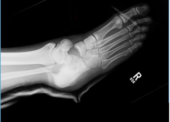
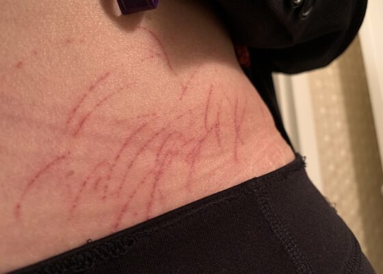
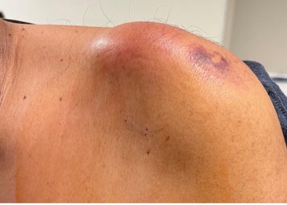
Examination of the right ankle demonstrated a large deformity of the superior talus with bruising and blanching of the overlying skin in the area of the Achilles tendon (see images 2,3). The remaining bones of the foot were not tender to palpation and the foot was neurovascularly intact throughout with only mild numbness in the area of the tented skin. Completing the trauma exam, the patient had no signs of head injury and no midline spinal tenderness to palpation. Inspection of the remaining long bones and joints showed no other injuries. There were mild skin scrapes on the right flank from the fall. X-rays of the right foot and ankle showed a longitudinal fracture of the calcaneal tuberosity from the articular surface to the posterior surface (see red outline) with extension into the subtalar joint (blue lines) and roughly 1.8 cm displacement between the fracture segments (yellow double arrow). These findings represented a tongue-type calcaneal bone fracture.
OrthopedicsTraumaVisual EM
Creative Commons images

Mushroom for Improvement Case Report: The Importance of Involving Mycologists

Gary Bhagat, MD*, Marit Tweet, MD^ and Steven Aks, MD^†

DOI: https://doi.org/10.21980/J8ZW7W Issue 7:4 No ratings yet.
The mushroom displayed here is large and lacks any gills. Small puffball mushrooms can resemble young immature button top Amanita type mushrooms. Opening the Amanita mushroom should reveal apparent gills and quickly differentiate the two- -the puffball mushroom should have a white interior without gills.
Abdominal/GastroenterologyToxicologyVisual EM
Creative Commons images

Evaluation of ACE-inhibitor Induced Laryngeal Edema Using Fiberoptic Scope: A Case Report

Joya Singh, DO* and Colin Danko, MD *

DOI: https://doi.org/10.21980/J83P9T Issue 7:3 No ratings yet.
Physical exam was initially significant for swelling isolated to the right sided cheek and upper lip. There was no edema to lower lip, uvular swelling, or swelling to the submandibular space. She was speaking full sentences and did not endorse any voice changes. Initial vital signs were as follows: BP 125/77, HR 74, RR 16, and oxygen saturation of 100% on room air. Approximately 40 minutes later, after 125 mg solumedrol intravenous (IV) and 50mg diphenhydramine by mouth, swelling had spread to the entire upper lip and the patient reported spreading to her jaw (Photo 1). Although no jaw or submandibular edema was appreciated on physical exam, a flexible fiberoptic laryngoscope was used to evaluate the patient’s airways given worsening symptoms. Viscous lidocaine was applied intranasally five minutes prior to the procedure. The patient was positioned in a seated position on the stretcher. A flexible fiberoptic laryngoscope was then inserted through the nares and advanced slowly. Laryngoscopy showed diffuse edema of the epiglottis, arytenoids, and ventricular folds (see photos 2-4). Vital signs and respiratory status remained stable both during and after the procedure.
ENTProceduresVisual EM
Creative Commons images

A Case Report of Fournier’s Gangrene

Huy Alex Duong, BS*, Mark Slader, BS*, Jana Florian, MD* and Jonathan Smart, MD*

DOI: https://doi.org/10.21980/J8Z356 Issue 7:2 No ratings yet.
Physical exam revealed a comfortable-appearing male patient with tachycardia and a regular cardiac rhythm. The genitourinary exam indicated significant erythema and fluctuance of the bilateral lower buttocks with extension to the perineum. Black eschar and ecchymosis were also noted at the perineum. There was significant tenderness to palpation that extended beyond the borders of erythema. There was no palpable crepitus on initial examination. Physical exam was otherwise unremarkable.
Infectious DiseaseUrologyVisual EM
Creative Commons images

A Case Report of the Rapid Evaluation of a High-Pressure Injection Injury of a Finger Leading to Positive Outcomes

Nathaniel Hansen, MD* and Colin Danko, MD*

DOI: https://doi.org/10.21980/J8TD2X Issue 7:2 No ratings yet.
On exam the patient was noted to have a punctate wound to the ulnar aspect of his right index finger, just proximal to the distal interphalangeal joint. The finger appeared pale and taut, with absent capillary refill. The patient displayed diminished range of motion with both extension and flexion of the joints of the finger. Sensation was absent and no doppler flow was appreciated to the distal aspects of the finger. X-ray of the hand was obtained and showed many small foreign bodies in the soft tissue and extensive radiolucent material consistent with gas or oil-based material to the palmar aspect of the index finger tracking up to the level of the metacarpal heads.
OrthopedicsVisual EM
‹2›»
Page 2 of 13

JETem is an online, open access, peer-reviewed, journal-repository for EM educators. We are PMC Indexed.

Most Viewed

  • The Silent Saboteur: Teaching the Clinical Implications of Occult Hypoxemia & Social Determinants of Health via a Pulmonary Embolism Case
  • Diabetic Ketoacidosis and Necrotizing Soft Tissue Infection
  • My Broken Heart
  • Stabilization of Cardiogenic Shock for Critical Care Transport, a Simulation
  • Innovative Ultrasound-Guided Erector Spinae Plane Nerve Block Model for Training Emergency Medicine Physicians

Visit Our Collaborators

Creative Commons Licence
This work is licensed under a Creative Commons Attribution 4.0 International License.

About

Education

Learners should benefit from active learning. JETem accepts submissions of team-based learning, small group learning, simulation, podcasts, lectures, innovations, curricula, question sets, and visualEM.

Scholarship

We believe educators should advance through the scholarship of their educational work. JETem gives educators the opportunity to publish scholarly academic work so that it may be widely distributed, thereby increasing the significance of their results.

Links

  • Home
  • Aim and Scope
  • Current Issue
  • For Reviewers
  • Instructions for Authors
  • Contact Us

Newsletter

Sign up to receive updates from JETem regarding newly published issues and findings.

Copyright Creative Commons Attribution 4.0 International