Issue 6:2
A Low-Cost Facial and Dental Nerve Regional Anesthesia Task Trainer
DOI: https://doi.org/10.21980/J8RP9QBy the end of this educational session, learners should be able to: 1) describe and identify relevant anatomy for supra-orbital, infra-orbital, mental, and inferior alveolar nerves and 2) successfully demonstrate supra-orbital, infra-orbital, mental, and inferior alveolar nerve blocks using a partial task trainer.
Teaching about the Wild: A Survival Course as a Novel Resident Educational Experience
DOI: https://doi.org/10.21980/J8N06RBy the end of the session the learner will be able to: 1) differentiate at least three different methods for water purification 2) describe how to erect a temporary survival shelter 3) construct a survival pack for personal use emphasizing multi-use items 4) demonstrate how to make a fire without a direct flame supply.
Escape the EM Boards: Interactive Virtual Escape Room for GI Board Review
DOI: https://doi.org/10.21980/J8H63FBy the end of this didactic activity, learners will be able to: 1) identify causes of upper gastrointestinal bleeding; 2) recall test-taking buzzwords for infectious causes of diarrhea; 2) acknowledge the correct hepatitis B titers that correspond with various clinical scenarios; 3) describe the management for alkali caustic ingestions; 4) determine the components of Maddrey Discriminant Function Score, Charcot’s triad, Ranson’s Criteria for Pancreatitis, and Glasgow-Blatchford Score; 5) diagnose specific gastrointestinal diseases from a clinical description; 6) choose the correct gastrointestinal diagnosis based on clinical image findings; 7) demonstrate teamwork in solving problems.
Let’s Escape Didactics: Virtual Escape Room as a Didactic Modality in Residency
DOI: https://doi.org/10.21980/J8CH2XBy the end of the activity, learners should be able to: 1) identify the hazardous chemicals associated with house fires; 2) classify burn injury according to depth, extent and severity based on established standards; 3) recall the actions to take in response to fire emergencies (R.A.C.E. and P.A.S.S. acronyms); 4) recall key laboratory features of cyanide and carbon monoxide poisonings; 5) identify appropriate management strategies for smoke inhalation injuries; 6) recite the treatment for cyanide and carbon monoxide poisonings; 7) describe the management of the burn injuries; 8) communicate and collaborate as a team to arrive at solutions of problems; 9) display task-switching and leadership skills during exercise; and 10) evaluate virtual escape room experience.
An Observation Medicine Curriculum for Emergency Medicine Education
DOI: https://doi.org/10.21980/J87P92The primary goal of this observation medicine curriculum is to train current EM residents in short-term acute care beyond the initial ED visit. This entails caring for patients from the time of their arrival to the OU to the point when a final disposition from the OU is determined, be it inpatient admission or discharge to home.
A Pediatric Emergency Medicine Refresher Course for Generalist Healthcare Providers in Belize: Respiratory Emergencies
DOI: https://doi.org/10.21980/J84063This curriculum presents a refresher course in recognizing and stabilizing pediatric acute respiratory complaints for generalist healthcare providers practicing in LMICs. Our goal is to implement this curriculum in the small LMIC of Belize. This module focuses on common respiratory complaints, including asthma, bronchiolitis, pneumonia and acute airway management.
Point of Care Ultrasound as a Diagnostic Tool to Detect Small Bowel Obstruction in the Emergency Department: A Case Report
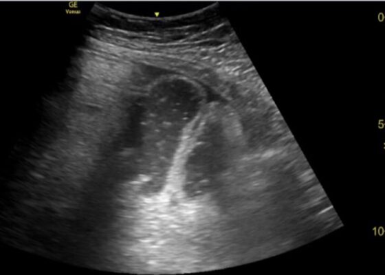
DOI: https://doi.org/10.21980/J8XD1GThe ultrasound findings suggestive of small bowel obstruction (SBO) are typically visualized in video; however, certain still images can also demonstrate SBO including greater than three dilated loops of small bowel (>2.5 cm), thickened-walled bowel (>3 mm), visualization of plicae circulares, and extraluminal fluid caused by inflammatory changes along the bowel wall, which are all highly suggestive of SBO.3
In this patient’s case, we were able to visualize several dilated loops of small bowel (red arrow) with oscillating intraluminal contents known as “Whirl Sign.” Additionally, we were able to visualize extraluminal fluid, demonstrated as an anechoic triangular-shaped collection. The characteristic shape of this triangular shaped collection of fluid is known as a “Tanga Sign,” given its name due to way it looks similar to the lower half of a bikini (blue arrow). Tanga sign can occur when the loops of dilated bowel appear prominent in contrast to the inflammatory extraluminal fluid in an SBO. These ultrasound findings were highly concerning for SBO which was later confirmed on CT imaging of the abdomen, which demonstrated SBO with a transition point in the left lower quadrant.
Case Report: Altered with PRES
DOI: https://doi.org/10.21980/J8NW73On vitals, the patient was found to be consistently hypertensive to the 230s/160s. Point-of-care glucose was within normal limits. Noncontrast CT imaging of the head revealed no acute intracranial hemorrhage or evidence of ischemic stroke, but was remarkable for areas of biparietal subcortical low-attenuation (white arrows), concerning for PRES. Patient subsequently underwent CT angiogram imaging of the head with perfusion which revealed no large vessel occlusion.
1›
Page 1 of 2


