• Registration
  • Login
JETem
  • Home
  • About
    • Aim and Scope
    • Our Team
    • Editorial Board
    • FAQ
  • Issues
    • Current Issue
    • Ahead of Print
    • Past Issues
  • Visual EM
    • Latest Visual EM
    • Search Visual EM
    • Thumbnail Library
  • For Authors
    • Instructions for Authors
    • Submit to JETem
    • Photo Consent
    • Policies
      • Peer Review Policy
      • Copyright Policy
      • Editorial Policy, Ethics and Responsibilities
      • Conflicts of Interest & Informed Consent
      • Open Access Policy
  • For Reviewers
    • Instructions for JETem Reviewers
    • Interested in Being a JETem Reviewer?
  • Topic
    • Abdominal / Gastroenterology
    • Administration
    • Board Review
    • Cardiology / Vascular
    • Clinical Informatics, Telehealth and Technology
    • Dermatology
    • EMS
    • Endocrine
    • ENT
    • Faculty Development
    • Genitourinary
    • Geriatrics
    • Hematology / Oncology
    • Infectious Disease
    • Miscellaneous
    • Neurology
    • Ob / Gyn
    • Ophthalmology
    • Orthopedics
    • Pediatrics
    • Pharmacology
    • Procedures
    • Psychiatry
    • Renal / Electrolytes
    • Respiratory
    • Social Determinants of Health
    • Toxicology
    • Trauma
    • Ultrasound
    • Urology
    • Wellness
    • Wilderness
  • Modality
    • Curricula
    • Innovations
    • Lectures
    • Oral Boards
      • Structured Interview
      • Communication Case
    • Podcasts
    • Simulation
    • Small Group Learning
    • Team Based Learning
    • Visual EM
  • Contact Us

Issue 3:1

Creative Commons images

A Sublingual Mass in a 5-Year-Old Male

Chris A Rees MD, MPH*, Andrea T Cruz, MD, MPH^ and Corrie E Chumpitazi, MD, MS^

DOI: https://doi.org/10.21980/J8H622Issue 3:1 No ratings yet.
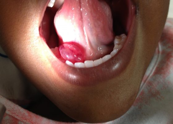
Oral ranula. On oral inspection he had a 3 x 1.5 cm, soft, non-tender, mobile, cystic, sublingual mass on the right aspect of the floor of his mouth that did not move with swallowing (Image 1). There was mild associated submandibular swelling on the right side of his face.
ENTVisual EM
Creative Commons images

Pneumomediastinum After Cervical Stab Wound

Chad Correa, BS* and Emily Ma, MD^

DOI: https://doi.org/10.21980/J87P79 Issue 3:1 No ratings yet.
Anteroposterior (AP) chest X-ray showed subcutaneous emphysema of the neck, surrounding the trachea (red arrows), right side greater than left, and a streak of gas adjacent to the aortic arch (white arrow). Computed tomography angiogram (CTA) of the neck showed air outside of the trachea, positive for pneumomediastinum (blue arrows). 
TraumaRespiratoryVisual EM
Creative Commons images

A Case of Acute Cholecystitis

Chad Correa, BS* and Lindsey Spiegelman, MD^

DOI: https://doi.org/10.21980/J8405QIssue 3:1 No ratings yet.
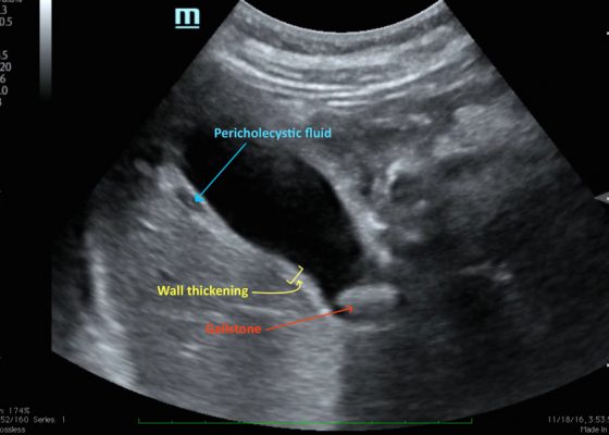
Bedside point-of-care ultrasound revealed a distended gallbladder, thickened gallbladder wall, pericholecystic fluid, and a stone in the neck of the gallbladder indicative of acute cholecystitis.
Abdominal/GastroenterologyVisual EM
Creative Commons images

Computed Tomography and Ultrasound Diagnosis of Spontaneous Subcapsular Renal Hematoma

Samer Assaf, MD*, Thalia Nguyen* and Alisa Wray, MD*

DOI: https://doi.org/10.21980/J8062DIssue 3:1 No ratings yet.
Bedside ultrasound was performed and demonstrated a hypoechoic area within the left kidney (images not shown). The non-contrast computed tomography (CT) of the abdomen and pelvis shows a significantly enlarged left kidney and a region of high-attenuation encapsulating the left kidney, concerning for acute hemorrhage.
Renal/ElectrolytesVisual EM
Creative Commons images

Point-of-care Ultrasound for the Diagnosis of a Gluteal Abscess

Kevin Roy, BS*, Robert Rowe, MD* and Maili Alvarado, MD^

DOI: https://doi.org/10.21980/J8VH1W Issue 3:1 No ratings yet.
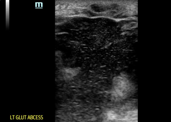
POCUS reveals a large, hypoechoic soft tissue abscess with debris and tracks extending to the bottom of the image. Furthermore, when compressed, movement of the abscess contents is appreciated. There is also superficial cobble-stoning consistent with overlying cellulitis and soft tissue edema.
UltrasoundInfectious DiseaseVisual EM
Creative Commons images

Perianal Abscess

Luke Walls-Smith, BS* and Jonathan Peña, MD*

DOI: https://doi.org/10.21980/J8QP81Issue 3:1 No ratings yet.
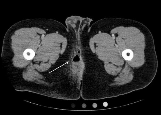
Computed Tomography (CT) of the Pelvis with intravenous (IV) contrast revealed a 5.7 cm x 2.4 cm air-fluid collection in the right perianal soft tissue along the right gluteal cleft, with surrounding fat stranding, consistent with a perianal abscess with cellulitis.
Abdominal/GastroenterologyInfectious DiseaseVisual EM
Creative Commons images

Tricuspid Annular Plane Systolic Excursion (TAPSE) in a Patient with Pulmonary Emboli

Nicole Zawada, BS*, Ethan Kunstadt, MD* and Maili Alvarado, MD^

DOI: https://doi.org/10.21980/J8M052 Issue 3:1 No ratings yet.
Video 1 and Image 1 show a thrombus in the patient’s IVC. Video 2 and Images 2-3 demonstrate a positive TAPSE of less than 17mm (blue arrow length) with a significantly dilated RV, indicating abnormal excursion of the tricuspid annulus and right ventricular dysfunction.  
RespiratoryCardiology/VascularVisual EM
Creative Commons images

Point-of-care Ultrasound Detection of Endophthalmitis

James Tucker MD, PhD*, Jonathan Patane, MD^ and Shadi Lahham, MD, MS^

DOI: https://doi.org/10.21980/J8G634 Issue 3:1 No ratings yet.
The patient’s ultrasound revealed an attached retina and a complex network of hyperechoic, mobile, membranous material in the posterior segment.
OphthalmologyUltrasoundVisual EM
Creative Commons images

Acute Pyelonephritis with Perinephric Stranding on CT

Ronald Goubert* and Jonathan Peña, MD*

DOI: https://doi.org/10.21980/J8BH0VIssue 3:1 No ratings yet.
A CT abdomen and pelvis with IV contrast showed neither nephrolithiasis nor diverticulitis, and instead showed heterogeneous enhancement of the left kidney with mild edematous enlargement and striated left nephrogram. Significant perinephric stranding (red arrows) was also noted and was consistent with severe acute pyelonephritis.
Renal/ElectrolytesInfectious DiseaseVisual EM
Creative Commons images

Empyema

Jonathan Patane, MD* and Daniel Ritter, BS^

DOI: https://doi.org/10.21980/J86P9RIssue 3:1 No ratings yet.
The chest X-ray shows a large fluid collection in the right lung demonstrated by the opacification that blunts the costophrenic angle on the right side. There is also a meniscus present, which is generally indicative of fluid.  Chest computed tomography (CT) demonstrated an infiltrate with a mixture of densities within the same collection, consistent with a loculated effusion and concerning for an empyema.
RespiratoryInfectious DiseaseVisual EM
‹2
Page 2 of 2

JETem is an online, open access, peer-reviewed, journal-repository for EM educators. We are PMC Indexed.

Most Viewed

  • The Silent Saboteur: Teaching the Clinical Implications of Occult Hypoxemia & Social Determinants of Health via a Pulmonary Embolism Case
  • Diabetic Ketoacidosis and Necrotizing Soft Tissue Infection
  • My Broken Heart
  • Stabilization of Cardiogenic Shock for Critical Care Transport, a Simulation
  • Innovative Ultrasound-Guided Erector Spinae Plane Nerve Block Model for Training Emergency Medicine Physicians

Visit Our Collaborators

Creative Commons Licence
This work is licensed under a Creative Commons Attribution 4.0 International License.

About

Education

Learners should benefit from active learning. JETem accepts submissions of team-based learning, small group learning, simulation, podcasts, lectures, innovations, curricula, question sets, and visualEM.

Scholarship

We believe educators should advance through the scholarship of their educational work. JETem gives educators the opportunity to publish scholarly academic work so that it may be widely distributed, thereby increasing the significance of their results.

Links

  • Home
  • Aim and Scope
  • Current Issue
  • For Reviewers
  • Instructions for Authors
  • Contact Us

Newsletter

Sign up to receive updates from JETem regarding newly published issues and findings.

Copyright Creative Commons Attribution 4.0 International