CT
Case Report: Thoracic Aortic Dissection in a Previously Healthy Male with an Unusual Inciting Factor
DOI: https://doi.org/10.21980/J8G92SA non-contrast computed tomography (CT) scan was negative for a suspected ureteral stone. However, there were aortic calcifications visualized at the infrarenal level that were notable given the patient’s age (red arrow). Given this finding in conjunction with the patient’s symptoms, bedside transabdominal ultrasonography was performed which revealed an intraluminal echogenic flap within the aorta near the common iliac arteries. CT angiography (CTA) with delayed contrast protocol revealed an extensive Stanford type A aortic dissection with involvement of the aortic root (purple arrow), brachiocephalic trunk, ostia of the left subclavian artery, descending aorta (blue arrow), bilateral common iliac arteries, and left internal iliac artery.
Case Report: Altered with PRES
DOI: https://doi.org/10.21980/J8NW73On vitals, the patient was found to be consistently hypertensive to the 230s/160s. Point-of-care glucose was within normal limits. Noncontrast CT imaging of the head revealed no acute intracranial hemorrhage or evidence of ischemic stroke, but was remarkable for areas of biparietal subcortical low-attenuation (white arrows), concerning for PRES. Patient subsequently underwent CT angiogram imaging of the head with perfusion which revealed no large vessel occlusion.
A Case Report of Cardiac Tamponade
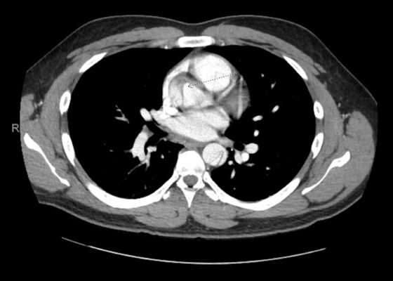
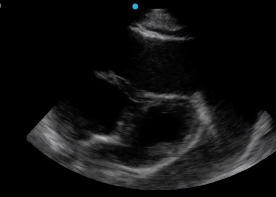
DOI: https://doi.org/10.21980/J8J644The patient was in noticeable respiratory distress and had oxygen saturation of 94% on room air. Bilateral jugular venous distention with severe right supraclavicular lymphadenopathy and diffuse bilateral wheezing was present. Although muffled heart sounds and hypotension are part of Beck’s Triad, these were not present in this case. Electrocardiogram obtained on arrival showed a sinus tachycardia with low-voltage QRS complexes and electrical alternans. Low voltage QRS can be seen on the ECG provided and is demonstrated by the low amplitude of the QRS complexes seen on all the leads. Electrical alternans may have an alternating axis or amplitudes of the QRS complex. Alternating axis is best visualized in V4-V6 on this ECG while alternating amplitudes are seen throughout the rest of the ECG. Computed tomography angiogram (CTA) of the chest revealed a large pericardial effusion with bilateral pulmonary emboli and a right upper lobe mass. A bedside transthoracic echocardiogram (TTE) was then performed and confirmed the large effusion, but also showed right ventricular collapse during diastole, indicative of cardiac tamponade.
A Different Type of Tension Headache: A Case Report of Traumatic Tension Pneumocephalus
DOI: https://doi.org/10.21980/J8DH0GCT head without contrast demonstrated a minimally displaced fracture of the frontal sinuses at the midline underlying his known laceration that involved the anterior and posterior tables of the calvarium. This is seen on the sagittal view and indicated by the blue arrow. There was a small volume of underlying subarachnoid hemorrhage along the falx. There was also extensive pneumocephalus most pronounced along the bilateral anterior frontal convexity associated with the frontal sinus fracture, seen on the axial image and indicated by the red arrow. This pattern of air is commonly referred to as the “Mount Fuji” sign.6 Other intracranial air can also be seen on the sagittal image and is indicated by the white arrow.
Jefferson Fracture and the Classification System for Atlas Fractures, A Case Report
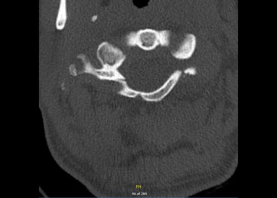
DOI: https://doi.org/10.21980/J88P9CComputed tomography (CT) revealed a burst fracture (Jefferson) of the anterior arch (white arrows) and of the posterior arch (yellow arrows) of the first cervical vertebrae (C1). There was also a fracture of the right lateral mass (blue arrow) of C1 with mild lateral subluxation of the lateral masses (curved arrows).
Using Point-of-Care Ultrasound to Expedite Diagnosis of Necrotizing Fasciitis: A Case Report
DOI: https://doi.org/10.21980/J85051A consultative scrotal ultrasound was performed, which was read as showing a small right hydrocele, small bilateral scrotal pearls, and normal-appearing testes. Although present, there was no mention of subcutaneous air suggestive of NF, seen in figure 1 as punctate hyperechoic foci (arrowhead) with ring-down artifact known as dirty shadowing (arrow). Also, subcutaneous thickening (asterisk) and free fluid (arrow) were seen as shown in figure 2, although their clinical relevance was not recognized in the radiologist's final report. Figure 3 shows an abdominal and pelvic CT that re-demonstrates subcutaneous air in the scrotum and lower abdomen (arrow) as well as fascial thickening of the perineum and free intra-abdominal air. After these images, the patient was transferred to our hospital for further management. Almost immediately after the patient's arrival, POCUS was employed. As seen in figures 4, we were able to identify in just a few minutes extensive subcutaneous air accompanied by dirty shadowing, as well as re-demonstration of subcutaneous thickening, fluid collections, and a right hydrocele. Even without the outside hospital's CT, the sonographic findings were highly suggestive for the diagnosis of NF of the perineum, also known as Fournier’s gangrene.
A Case Report on Detecting Porcelain Gallbladder form Wall-Echo-Shadow Sign on Point-of-Care Ultrasound
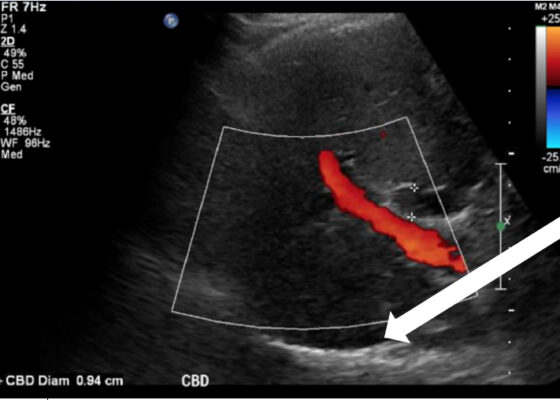
DOI: https://doi.org/10.21980/J8164GPoint-of-care ultrasound (POCUS) was performed by the emergency physician. Gallbladder ultrasound (US) should be performed using a curvilinear probe. If the patient’s body habitus does not allow for the use of a curvilinear probe, a phased array probe may be used. To find the gallbladder with ultrasonography, two approaches are commonly used. Many physicians prefer the “subcostal sweep” in which the probe is placed on the xiphoid process in a sagittal plane and swept along the inferior costal margin until the gallbladder is visualized. If this does not adequately locate the gallbladder, the “X minus 7” approach may be used. In this approach, the probe is placed on the xiphoid (X) process in a transverse view and moved 7 centimeters (minus 7) to the patient’s right. This technique is useful for patients with a larger body habitus. If the gallbladder is still not visualized, placing the patient in left lateral decubitus position or asking them to take a deep breath and hold may help the ultrasonographer locate the gallbladder. The US revealed mild hepatic biliary duct dilation with cholelithiasis and sludge, but no additional evidence to suggest cholecystitis. The US image showed a dilated common bile duct at 0.94 cm and calcifications. Visualization of the gallbladder wall is essential in differentiating between a positive wall-echo-shadow (WES) sign and a porcelain gallbladder. While a hypoechoic gallbladder wall is indicative of a WES sign, a hyperechoic wall layer will indicate a calcified gallbladder wall, suggesting a porcelain gallbladder. In image 1, the hyperechoic gallbladder wall can be visualized (white arrow), suggesting the presence of porcelain gallbladder and distinguishing it from a positive WES sign.
Pulmonary Arteriovenous Malformation in a Patient with Suspected Hereditary Hemorrhagic Telangiectasia: A Case Report
DOI: https://doi.org/10.21980/J8M353Initial vital signs were unremarkable, including oxygen saturation of 98% on room air. The patient did not exhibit any signs of respiratory distress, and the lungs were clear to auscultation bilaterally. Labs were obtained, which showed normal hemoglobin at 15.8. Computed tomography (CT) of the chest (Video 1) showed a large left upper lobe arteriovenous malformation (AVM) with large feeding arteries and tortuous dilated draining veins (red arrow) measuring up to 3.8cm. Imaging also demonstrated nonspecific multifocal ground-glass opacities, which may have represented pulmonary hemorrhage (blue outline) from AVM without evidence of contrast extravasation to suggest active bleeding.


