Search By Modality
Found 645 Unique Results
Page 2 of 65
Page 2 of 65
Page 2 of 65
Case Report of Incarcerated Gastric Volvulus and Splenic Herniation in Undiagnosed Congenital Diaphragmatic Hernia in an Infant
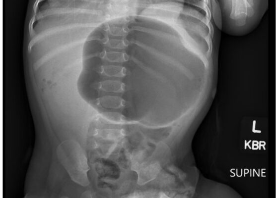
DOI: https://doi.org/10.21980/J8VD27An upper gastrointestinal series (UGI) showed an enteric tube with its tip in the stomach and side-port in the esophagus. There was a large amount of air in the stomach and a small volume of scattered distal bowel gas. The tip of an enteric tube was seen in the stomach (red arrow). Contrast partially filled the stomach, and the greater curvature was visualized superior to the lesser curvature in the left upper quadrant (blue arrow). The body of the stomach was herniated into the right chest through a Bochdalek hernia (blue star). There was a large amount of air in the stomach and a small volume of scattered distal bowel gas. These findings were consistent with mesenteroaxial gastric volvulus.
Beware of the Pediatric Limp: A Case of Mycoplasma Associated Acute Transverse Myelitis
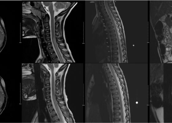
DOI: https://doi.org/10.21980/J8QQ1QAn MRI with contrast, T2 sequence was performed. In Figures a-d, the MRI of the patient’s brain and spinal cord on admission shows abnormal signals in the patient’s pons (lack of symmetrical gray-white differentiation on cross-section) along with hyperintensity (sagittally shown as brightness in what should be homogenously intense spinal cord) and significant central cord edema (with swelling seen as increased width) starting from C5 and continuing to the conus medullaris around L1/L2.
A Case Report of Calciphylaxis
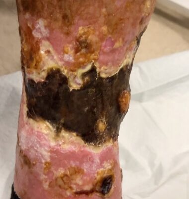
DOI: https://doi.org/10.21980/J8KW8VOn arrival for this visit, the patient was nontoxic appearing with stable vital signs. The physical exam was notable for deep, ulcerated, bilateral anterior leg wounds with purulent drainage and large areas of eschar (see photographs).
Case Report: Iatrogenic Bowel Perforation Following Dental Procedure
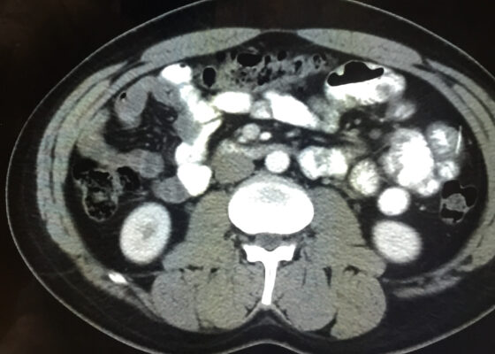
DOI: https://doi.org/10.21980/J8CD38The patient’s abdominal CT demonstrated a metallic foreign body in the left side of the abdomen within the small bowel, without surrounding induration or abscess. Radiology questioned whether the metallic foreign object perforated the bowel. Seen in the cross-sectional CT image, there is a hyperdense linear structure transversing the small intestinal wall, given that a portion of the structure was located outside of the lumen of the bowel.
Diabetic Ketoacidosis and Necrotizing Soft Tissue Infection
DOI: https://doi.org/10.21980/J89M0KAt the end of this oral board session, examinees will: 1) Demonstrate the ability to obtain a complete medical history and physical exam. 2) Identify and appropriately treat DKA. 3) Identify, treat, and make appropriate consults for NSTI. 4) Demonstrate effective communication of the treatment plan with the patient.
My Broken Heart
DOI: https://doi.org/10.21980/J85W7RBy the end of this simulation session, learners will be able to: 1) assess the hemodynamics of an LVAD patient by using a Doppler to determine mean arterial pressure, 2) Manage an arrhythmia in an LVAD patient with a suction event by addressing preload, 3) Identify and treat the source of hypovolemia (a massive lower gastrointestinal hemorrhage), 4) Perform clear closed-loop communication with other team members.
Stabilization of Cardiogenic Shock for Critical Care Transport, a Simulation
DOI: https://doi.org/10.21980/J82354ABSTRACT: Audience: This simulation is designed for critical care transport providers but can be easily adapted for the inpatient setting. It is applicable to an interdisciplinary team including nurses, respiratory therapists, medical students, emergency medicine residents, and emergency medicine attendings. Introduction: Cardiogenic shock carries an incredibly high burden of morbidity and mortality. Acute myocardial infarction accounts for 81% of cardiogenic
Innovative Ultrasound-Guided Erector Spinae Plane Nerve Block Model for Training Emergency Medicine Physicians
DOI: https://doi.org/10.21980/J8PW7DThis innovation model is designed to facilitate hands-on training of the ultrasound-guided ESP nerve block using a practical, realistic, and cost-effective ballistics gel model. By the end of this training session, learners should be able to: 1) identify relevant sonoanatomy on the created simulation model; 2) demonstrate proper in-plane technique; and 3) successfully replicate the procedure on a different target on the created training model.
Orthopaedic Surgery Didactic Session Improves Confidence in Distal Radius Fracture Management by Emergency Medicine Residents
DOI: https://doi.org/10.21980/J8K365By the end of this didactic session, learners should be able to: 1) assess DRF displacement on pre-reduction radiography and formulate reduction strategies, 2) perform a closed reduction of a DRF, 3) apply a safe and appropriate plaster splint to patient with a DRF and assess the patient’s neurovascular status, 4) assess DRF post-reduction radiography for relative fracture alignment, and 5) understand appropriate follow-up and necessary return precautions.


