
Visual EM

A Case Report of Granulomatosis with Polyangiitis Presenting with Chronic Conjunctivitis
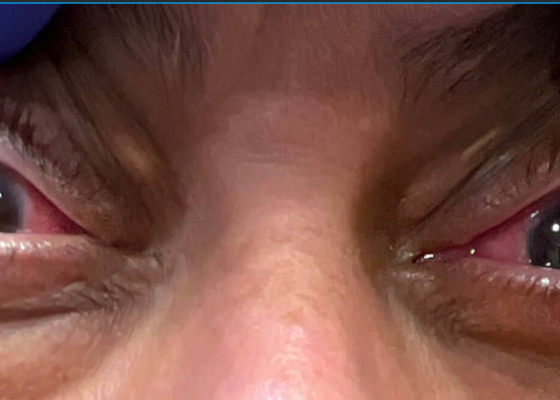
DOI: https://doi.org/10.5070/M5.52284Notable bilateral proptosis with conjunctival injection and limbic sparing (clear conjunctiva surrounding the margin of the cornea, shown by the arrow in the image of the eyes) was present.
Caught in the Whirl: A Case Report of Midgut Volvulus in an Elderly Patient
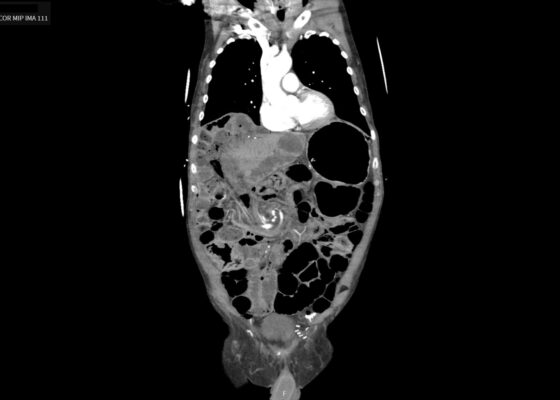
DOI: https://doi.org/10.5070/M5.52361An upright chest x-ray demonstrated air underneath the diaphragm bilaterally (yellow arrows), enhancing the diaphragmatic contour. A computed tomography angiography (CTA) abdomen and pelvis showed scattered pneumoperitoneum (red arrow), dilated segments of small bowel (orange arrow), and clockwise swirling of the mesentery (blue arrow), concerning for mesenteric volvulus with possible bowel perforation.
A Case Report of Acute Appendicitis Complicated by Appendicoliths
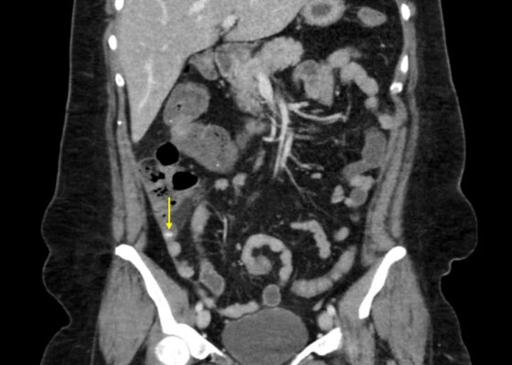
DOI: https://doi.org/10.5070/M5.52213Contrast-enhanced CT of the abdomen and pelvis was obtained. Coronal CT demonstrated a dilated appendix measuring up to 1.2 cm in diameter with multiple appendicoliths (yellow arrows), the largest measuring 1.1 cm. Mild periappendiceal fat stranding was present, consistent with acute appendicitis complicated by appendicoliths. The surrounding bowel and colon were normal in caliber and distribution. An additional coronal view demonstrated a dilated appendix (yellow arrow) containing an appendicolith with periappendiceal inflammatory changes. No evidence of perforation, abscess, or drainable fluid collection was identified.
Case Report: Acute Dyspnea in a Young Female
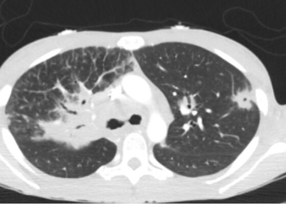
DOI: https://doi.org/10.5070/M5.52254The patient underwent computer tomography (CT) imaging of the chest, abdomen, and pelvis. Computer tomography of the chest showed bilateral diffuse pulmonary nodules in a perilymphatic distribution (yellow arrows), and diffusely increased mediastinal and hilar soft tissue densities (blue circle), likely representing lymphadenopathy; a left upper lobe lesion with central cavitation (orange circle) was also seen likely to be associated with the same disease process as the extensive lymphadenopathy. Imaging of the abdomen and pelvis was significant for diffuse hypodensities of the liver and spleen suggesting a multi-system process.
The Rash That Didn’t Blanch: A Case Report of Adult-Onset IgA Vasculitis with Underlying Cirrhosis and IgA Nephropathy
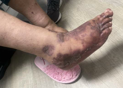
DOI: https://doi.org/10.5070/M5.52253Given the patient’s physical exam findings of tender palpable purpura to the lower extremities, dermatology was consulted for suspicion of acute small vessel vasculitis. A punch biopsy for hematoxylin and eosin (H&E) stain and direct immunofluorescence (DIF) was subsequently performed. The H&E demonstrated nonspecific purpura.
A Case Report on an Open Fracture Dislocation Injury of the Proximal Phalanx of the Thumb Resulting from Playing Cricket
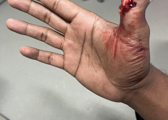
DOI: https://doi.org/10.5070/M5.52278There was an open injury to the volar aspect of the right thumb at the interphalangeal joint, with exposed bone. There was no active bleeding from the wound. He had intact sensation to the entire thumb and hand. His radial pulse was normal with normal capillary refill in all digits of his right hand. He had intact wrist flexion, extension, abduction, and adduction; however, he was unable to flex or extend his thumb secondary to the injury. He had no other injuries to the rest of his right upper extremity. An x-ray was obtained which showed a right thumb proximal phalanx intra-articular fracture (proximal fracture fragment outlined in yellow, distal fracture fragment outlined in pink) at the interphalangeal joint with dorsal dislocation of the distal phalanx (outlined in red). There were no radiopaque foreign bodies.
Woman with a Blackened Tongue: A Case Report
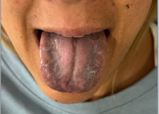
DOI: https://doi.org/10.5070/M5.52329The patient’s vital signs were blood pressure 109/72 mmHg, respiratory rate 18 breaths per minute, pulse 93 beats per minute, temperature 97°F. Physical examination revealed notable black discoloration of the tongue and gingiva, along with hyperpigmented skin exhibiting numerous diffuse nevi distributed across the face and upper extremities.
A Woman’s Infertility Journey Complicated by Severe Ovarian Hyperstimulation Syndrome – A Case Report
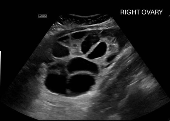
DOI: https://doi.org/10.5070/M5.52309Computed tomography (CT) of the abdomen and pelvis demonstrated pleural effusions in the lung bases (blue stars), ascites (blue stars), and enlarged ovaries with multiple cysts/follicles (white arrows). A formal pelvic US also demonstrated large volume ascites (white diamonds) and bilateral ovarian enlargement with numerous cysts. A CT angiogram (CTA) of the chest demonstrated small bilateral pleural effusions and no obvious pulmonary embolism.