Ultrasound
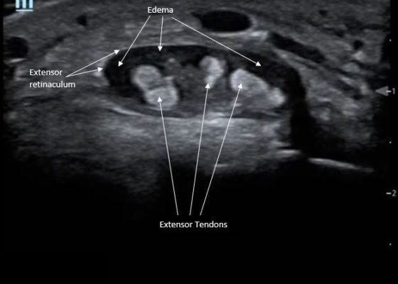
Point-Of-Care Ultrasound for the Diagnosis of Extensor Tenosynovitis
DOI: https://doi.org/10.21980/J8Q050Point-of-care ultrasound of the dorsal aspect of the left hand reveals a heterogenous hypoechoic fluid collection surrounding the extensor tendons (axial view) within the retinaculum consistent with edema. Longitudinal view shows anechoic fluid within the tenosynovium which is located between the anisotropic extensor tendon and linear hyperechoic synovial sheath. Longitudinal view also shows some cobblestoning, or tissue edema, superficial to the anisotropic extensor tendon. The patient’s contralateral right dorsal hand was scanned in a longitudinal view and shows no cobblestoning or hypoechoic fluid under the synovial sheath. The patient was diagnosed with tenosynovitis, and started on intravenous antibiotics.
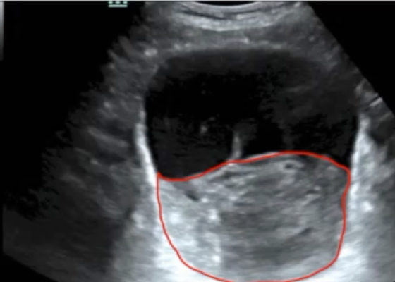
Point-of-care Ultrasound in the Diagnosis and Monitoring of Bladder Hematoma vs. Hemorrhage
DOI: https://doi.org/10.21980/J8092FBladder POCUS demonstrated 500mL of post void residual fluid, indicative of retention. Half of the volume is hyperechoic (red circle); this is likely the bladder wall hematoma. Could also consider sonographic artifact, bladder mass, or cystitis.1-2
Point-of-Care Ultrasound to Evaluate Intrahepatic Biliary Stent Function
DOI: https://doi.org/10.21980/J86S6NThe ultrasound image demonstrates severe intrahepatic biliary ductal dilatation without an obvious intrahepatic obstructive lesion, as pointed out by the white arrows. The hepatic vasculature is well-distinguished from the biliary tree via color flow doppler, as seen by the white arrowheads.
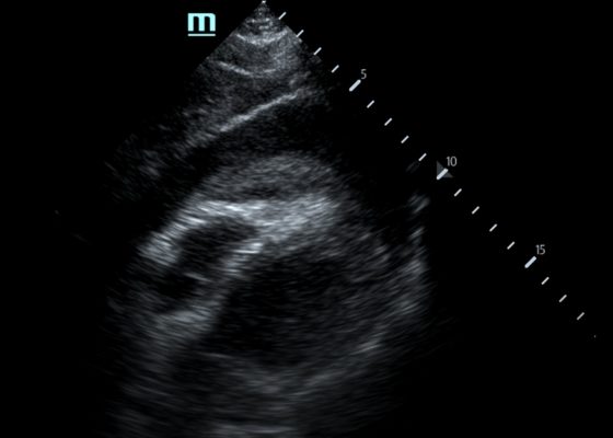
Pericardial Clot on Point-of-Care Ultrasound
DOI: https://doi.org/10.21980/J8ZH1TFocused assessment with sonography in trauma (FAST) scan was positive for a clinically significant pericardial effusion as evidenced by the hypoechoic fluid around the myocardium, indicated by the blue arrow in image 2. Findings are also consistent with tamponade process as evidenced by restricted expansion and collapse of the right ventricle during diastole. The hyperechoic floating structure between the pericardium and myocardium, adjacent to the right ventricle, represents a pericardial clot, indicated by the white arrow.The density of the pericardial clot differs from that of the myocardium, thus serving as an additional variable to avoid confusing this as part of the myocardial structure.
Radiolucent Foreign Body Seen on Point-of-Care Ultrasound but not on X-ray
DOI: https://doi.org/10.21980/J8WS77X-rays of the foot were obtained and no radiopaque foreign body was visualized. Due to high clinical suspicion for retained foreign body, a point-of-care ultrasound was performed by applying a high-frequency linear probe at the area of discomfort. In the long axis an ovoid focus of hypoechogenicity (orange outline) is visualized. Within this finding there is a linear focus (yellow line) of increased echogenicity measuring 1 mm in diameter and 1 cm in length. On short axis view, a rectangle focus (green dot) demonstrating shadowing (blue highlight) is seen.
Low-Cost, Low Fidelity Meat Model to Teach Ultrasound Guided Nerve Blocks
DOI: https://doi.org/10.21980/J83G9RUpon completion of this workshop, learners will be able to: 1) Describe the risks and benefits of ultrasound guided nerve blocks. 2) Choose the appropriate nerve to block based on the area that needs anesthesia. 3) Display proficiency in performing an ultrasound guided nerve block on meat models. 4) Verbalize confidence in successfully performing ultrasound guided regional anesthesia. By successfully meeting these objectives, we aim to improve learner confidence and clinical ability in performing ultrasound guided nerve blocks.
Point of Care Ultrasound Illustrating Small Bowel Obstruction
DOI: https://doi.org/10.21980/J8T637POCUS of the small bowel illustrated significantly dilated loops of bowel (white line), thickened bowel wall (white arrow) and to-and-fro peristalsis, consistent with small bowel obstruction.
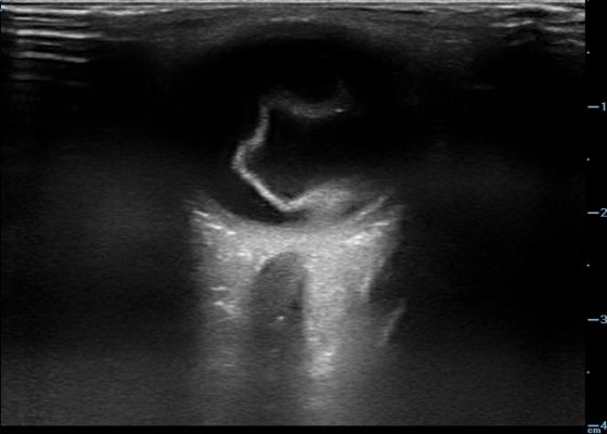
Retinal Detachment
DOI: https://doi.org/10.21980/J8204QBedside ocular ultrasound revealed a serpentine, hyperechoic membrane that appeared tethered to the optic disc posteriorly with hyperechoic material underneath. These findings are consistent with retinal detachment (RD) and associated retinal hemorrhage.


